

Monocyte-derived dendritic cells reflect the immune functional status of a chromophobe renal cell carcinoma patient: could it be a general phenomenon?
- PMID: 25314913
- PMCID: PMC11029287
- DOI: 10.1007/s00262-014-1625-9
Monocyte-derived dendritic cells reflect the immune functional status of a chromophobe renal cell carcinoma patient: could it be a general phenomenon?
Abstract
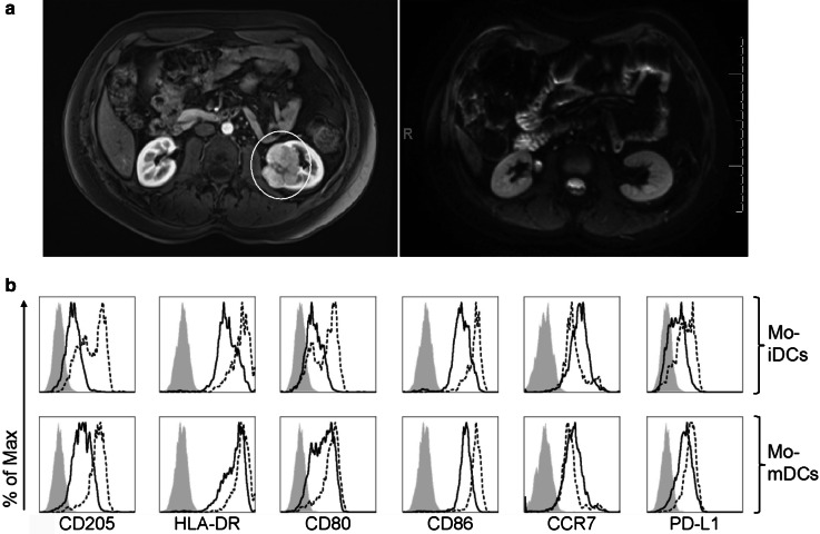
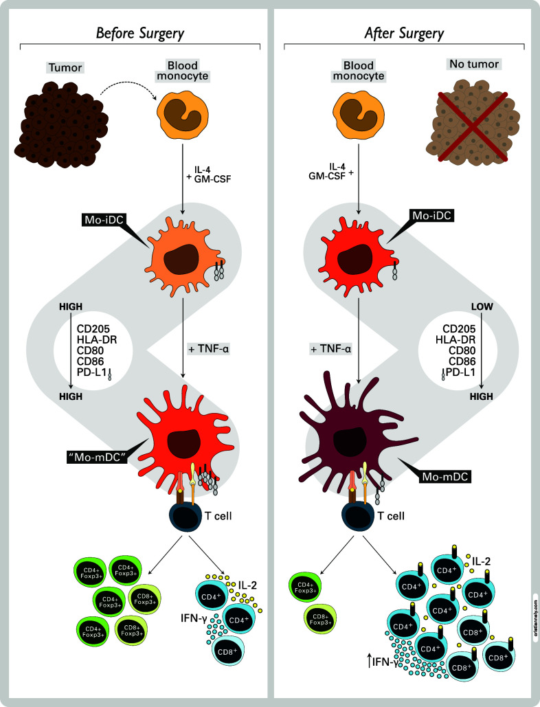
Purpose: The chromophobe renal cell carcinoma (ChRCC), though associated with a hereditary cancer syndrome, has a good prognosis after tumor removal. The lack of recurrence could be related to the absence of immune system compromise in patients or to an effective functional recovery of immune functions after tumor removal. Thus, we evaluated monocyte-derived dendritic cells (Mo-DCs) in a 34-year-old male who had a ChRCC, before and after tumor removal.
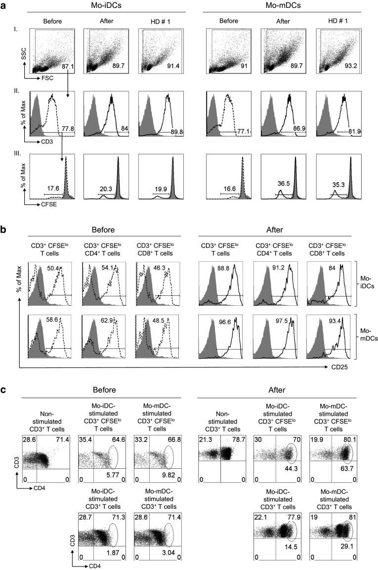
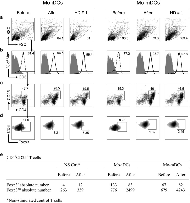
Methods: CD14(+) monocytes from the patient's peripheral blood, 1 week before and 3 months after partial nephrectomy, were differentiated in vitro into immature and mature Mo-DCs. These were harvested, analyzed by flow cytometry and used as stimulators of allogeneic T cells. Supernatants from cultures were collected for cytokine analysis.
Results: Tumor removal was associated with decreased expression of PD-L1, but also, surprisingly, of CD205, HLA-DR, CD80 and CD86 by Mo-DCs. Also, Mo-DC's ability to stimulate T cell proliferation increased, along with IL-2Rα expression and IFN-γ production. Simultaneously, the patients' Mo-DCs ability to induce Foxp3(+) T cells decreased after surgery. One-year postoperative follow-up shows no tumor recurrence.
Conclusion: The presence of a ChRCC affected Mo-DCs generated in vitro, which recovered their function after tumor removal. This indicates that the favorable outcome observed after ChRCC resection may be due to the restoration of immunocompetence. Furthermore, since functional alterations described for DCs within tumors may be also found in Mo-DCs, their accurate functional analysis-not restricted to the determination of their surface immunophenotype-may provide an indirect "window" to the tumor microenvironment.
Conflict of interest statement
The authors declare no financial or other conflict.
Figures
Similar articles
-
Inhibiting STAT5 by the BET bromodomain inhibitor JQ1 disrupts human dendritic cell maturation.J Immunol. 2015 Apr 1;194(7):3180-90. doi: 10.4049/jimmunol.1401635. Epub 2015 Feb 27. J Immunol. 2015. PMID: 25725100 Free PMC article.
-
1-alpha,25-Dihydroxyvitamin D3 (1,25(OH)(2)D(3)) hampers the maturation of fully active immature dendritic cells from monocytes.Eur J Endocrinol. 2001 Sep;145(3):351-7. doi: 10.1530/eje.0.1450351. Eur J Endocrinol. 2001. PMID: 11517017
-
Immunomodulatory dendritic cells generated from nonfractionated bulk peripheral blood mononuclear cell cultures induce growth of cytotoxic T cells against renal cell carcinoma.J Immunother. 2000 Jan;23(1):83-93. doi: 10.1097/00002371-200001000-00011. J Immunother. 2000. PMID: 10687141
-
Dendritic cells derived from metastatic cancer patients vaccinated with allogeneic dendritic cell-autologous tumor cell hybrids express more CD86 and induce higher levels of interferon-gamma in mixed lymphocyte reactions.Cancer Immunol Immunother. 2005 Jan;54(1):61-6. doi: 10.1007/s00262-004-0550-8. Cancer Immunol Immunother. 2005. PMID: 15693140 Free PMC article.
-
Immunosuppressive effect of renal cell carcinoma on phenotype and function of dendritic cells.Int Urol Nephrol. 2014 May;46(5):915-20. doi: 10.1007/s11255-013-0595-8. Epub 2013 Nov 8. Int Urol Nephrol. 2014. PMID: 24202958
Cited by
-
Tolerogenic IDO(+) Dendritic Cells Are Induced by PD-1-Expressing Mast Cells.Front Immunol. 2016 Jan 25;7:9. doi: 10.3389/fimmu.2016.00009. eCollection 2016. Front Immunol. 2016. PMID: 26834749 Free PMC article.
-
Human Dendritic Cells: Their Heterogeneity and Clinical Application Potential in Cancer Immunotherapy.Front Immunol. 2019 Jan 21;9:3176. doi: 10.3389/fimmu.2018.03176. eCollection 2018. Front Immunol. 2019. PMID: 30719026 Free PMC article. Review.
-
Pigmented Epithelioid Melanocytoma (Animal Types of Melanoma) on the Nose.Case Rep Oncol. 2018 Jun 13;11(2):378-382. doi: 10.1159/000490237. eCollection 2018 May-Aug. Case Rep Oncol. 2018. PMID: 30022939 Free PMC article.
-
The Therapeutic Potential of Tackling Tumor-Induced Dendritic Cell Dysfunction in Colorectal Cancer.Front Immunol. 2021 Oct 6;12:724883. doi: 10.3389/fimmu.2021.724883. eCollection 2021. Front Immunol. 2021. PMID: 34691029 Free PMC article. Review.
-
CD163+ tumor-associated macrophage accumulation in breast cancer patients reflects both local differentiation signals and systemic skewing of monocytes.Clin Transl Immunology. 2020 Feb 13;9(2):e1108. doi: 10.1002/cti2.1108. eCollection 2020. Clin Transl Immunology. 2020. PMID: 32082570 Free PMC article.
References
Publication types
MeSH terms
Substances
LinkOut - more resources
Full Text Sources
Other Literature Sources
Medical
Research Materials
