. 2014 Oct 14;7(347):ra97.
doi: 10.1126/scisignal.2005413.

Temporal and spatial regulation of epsin abundance and VEGFR3 signaling are required for lymphatic valve formation and function

Affiliations

Temporal and spatial regulation of epsin abundance and VEGFR3 signaling are required for lymphatic valve formation and function

Xiaolei Liu et al. Sci Signal. .

Abstract

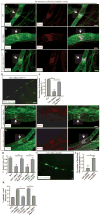
Lymphatic valves prevent the backflow of the lymph fluid and ensure proper lymphatic drainage throughout the body. Local accumulation of lymphatic fluid in tissues, a condition called lymphedema, is common in individuals with malformed lymphatic valves. The vascular endothelial growth factor receptor 3 (VEGFR3) is required for the development of lymphatic vascular system. The abundance of VEGFR3 in collecting lymphatic trunks is high before valve formation and, except at valve regions, decreases after valve formation. We found that in mesenteric lymphatics, the abundance of epsin 1 and 2, which are ubiquitin-binding adaptor proteins involved in endocytosis, was low at early stages of development. After lymphatic valve formation, the initiation of steady shear flow was associated with an increase in the abundance of epsin 1 and 2 in collecting lymphatic trunks, but not in valve regions. Epsin 1 and 2 bound to VEGFR3 and mediated the internalization and degradation of VEGFR3, resulting in termination of VEGFR3 signaling. Mice with lymphatic endothelial cell-specific deficiency of epsin 1 and 2 had dilated lymphatic capillaries, abnormally high VEGFR3 abundance in collecting lymphatics, immature lymphatic valves, and defective lymph drainage. Deletion of a single Vegfr3 allele or pharmacological suppression of VEGFR3 signaling restored normal lymphatic valve development and lymph drainage in epsin-deficient mice. Our findings establish a critical role for epsins in the temporal and spatial regulation of VEGFR3 abundance and signaling in collecting lymphatic trunks during lymphatic valve formation.


Conflict of interest statement

COMPETING INTERESTS: The authors declare that they have no competing interests.

Figures

Fig. 1
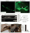
Fig. 1. Defective collecting lymphatic valve formation in LEC-DKO mice
A–H) Representative whole mount immunofluorescent staining of wild-type and LEC-DKO mesenteric lymphatic valves at E18.5 (A–D) and P6 (E–H) with antibodies against CD31, Prox1 and Integrin-α9. B, D and F, H are enlarged images of A, C and E, G. A, C: n=5 embryos per group; E, G: n=6 pups per group. Itgα9: Integrin-α9. V: vein, A: artery, L: collecting lymphatic. Scale bar, 100 μm (A, C, E, G); Scale bar, 50 μm (B, D, F, H). I–J) Representative whole mount immunofluorescent staining of wild-type (I) and LEC-DKO (J) mesenteric lymphatic valves at P13 with antibodies against CD31 (n=6 pups per group). Scale bar, 100 μm. K–N) Representative bright field images of chyle-filled mesenteric vessels in both wild-type (K, L) and LEC-DKO mice (M, N) (n=6 mice per group). LEC-DKO mice (L) have immature valve morphology (black arrow) and dilated vessels compared to wild-type mice (N). L: Lymphatic vessels. Scale bar, 1 mm (K–M); Scale bar, 50 μm (L–N). O) Quantification of percentages of V-shaped valve numbers of in wild-type and LEC-DKO mice at different developmental stages. *, P<0.05; geographic regression. All values are mean ± SD (n=3 independent experiments).
Fig. 2
Fig. 2. Impaired lymphatic drainage in LEC-DKO mice
A–B) Representative images of ear lymphatic drainage after FITC-Dextran injection in P14 wild-type (A) and LEC-DKO (B) mice (n=9 mice per group). Arrow indicates a defective valve. Scale bar, 50 μm. C) Representative fluorescence microlymphangiography of FITC-Dextran injection into tail tips of P14 wild-type (n=4) and LEC-DKO mice (n=6) analyzed in 3 independent experiments. Black arrow above the images marks the direction of lymphatic transport. Scale bar, 1 cm. Quantification of FITC-Dextran labeling intensity is shown at right. Two-way ANOVA. D) Representative images of popliteal lymph nodes (black arrows) after Evans blue injection into hind foot pads of P6 wild-type and LEC-DKO pups (n=6 pups per group analyzed in 3 independent experiments). White dashed lines indicate a possible collecting lymphatic vessel. Scale bar, 5 mm. Quantification of Evans blue intensity is shown at right. All values are mean ± SD. *p<0.05; Logistic regression.
Fig. 3
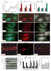
Fig. 3. Reciprocal expression of VEGFR3 and epsin in developing collecting lymphatic vessels
A) Quantification of qRT-PCR analysis of epsin 1 and epsin 2 mRNA in mesenteric lymphatics at developmental stages of E14, E16, E18, P0 and P6 (n=3 independent experiments). One-way ANOVA. B) Quantification of qRT-PCR analysis of epsin 1, epsin 2 and CX37 mRNA from mouse LECs pretreated with or without PP2 under steady shear stress or static conditions (n=3 independent experiments). One-way ANOVA. C) Quantification of qRT-PCR analysis of epsin 1, epsin 2 and CX37 mRNA from mouse LECs under laminar shear stress or static conditions (n=3 independent experiments). All values are mean ± SD. *p<0.05; Student t test. D–F) Representative immunostaining of mouse LECs after 16 hours of static, laminar shear stress or oscillatory shear stress conditions using antibodies against epsin 1, epsin 2 and counterstained with DAPI (n=3 independent experiments). Scale bar, 10 μm. White arrow indicates the colocalization of epsin 1 and epsin 2. G–I) Representative whole mount immunostaining of wild-type mesenteric collecting lymphatic vessels at developmental stages E16 (n=6 embryos) (G), P0 (n=5 pups) (H), and P6 (n=4 pups) (I) using antibodies against VEGFR3. Scale bar, 50 μm. J) Western blot analysis of epsin 1, epsin 2, VEGFR3, and p-VEGFR3 in isolated mesenteric LECs at developmental stages E14, E16, E18, P0 and P6. K) Quantification of epsin 1, epsin 2, and VEGFR3 protein abundance normalized to Tubulin (left panel) and VEGFR3 phosphorylation normalized to total VEGFR3 abundance (right panel) (n=3 independent experiments). All values are mean ± SD. *p<0.05 compared to E14; One-way ANOVA.
Fig. 4
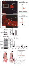
Fig. 4. Increased VEGFR3 abundance in LEC-DKO collecting lymphatic trunk LECs
A–B) Representative whole mount immunostaining of wild-type (A) and LEC-DKO (B) mesenteric collecting lymphatic vessels at P5 using antibodies against VEGFR3 (n=5 pups per group). White arrows indicate lymphatic valves. The diagrams at the right are models for how epsin 1 and 2 facilitate the decrease in VEGFR3 abundance in collecting lymphatic trunk LECs to ensure proper valve formation. Scale bar, 100 μm. C) Western blotting analysis of VEGFR3 abundance in skin LECs from WT and LEC-DKO mice. D) Quantification of VEGFR3 abundance normalized to Tubulin (n=3 independent experiments). E) Western blotting analysis of VEGFR3, VEGFR2, VEGFR1, EGFR, PDGFRβ, and TGFβR1 abundance in mesenteric LECs from WT and LEC-DKO mice. F) Quantification of receptor protein fold changes normalized to Tubulin (n=3 independent experiments). All values are mean ± SD. *p<0.05; One-way ANOVA. G) Schematic summary of the predicted roles of epsins in temporally and spatially regulating VEGFR3 abundance during lymphatic valve formation. Blue arrow indicates the two different types of lymph flow. Diagram at the right indicates the possible regulatory role of epsins which are increased by steady flow thus reducing VEGFR3 abundance to ensure proper valve formation.
Fig. 5
Fig. 5. Decreased ligand-induced VEGFR3 internalization and signaling termination in epsin-deficient LECs
A) FACS analysis of the surface abundance of VEGFR3 in LECs from wild-type or LEC-DKO mice. Quantification is shown at right (n=6 independent experiments). B) Western blot analysis of internalized biotinylated VEGFR3 detected by pull down with streptavidin beads and Western blotting with VEGFR3 antibodies. Quantification of VEGFR3 internalization is shown at right (n=4 independent experiments). C) Mouse LECs from wild-type and LEC-DKO mice were stimulated with VEGFC c156s for indicated time points and lysates were followed by immunoprecipitation with VEGFR3 antibodies or control IgG. Phospho-VEGFR3 signals were detected by Western blotting using 4G10 antibodies. D) Quantification of total and phosphorylated VEGFR3 shown in (C) (n=3 independent experiments). E) Mouse LECs from wild-type and LEC-DKO mice were stimulated with VEGFC c156s for the indicated time points, and VEGFR3 downstream signaling was analyzed by Western blotting with the indicated antibodies. F) Quantification of VEGFR3 downstream signaling shown in (E) (n=3 independent experiments). All values are mean ± SD. *p<0.05; **p<0.01; Two-way ANOVA.
Fig. 6
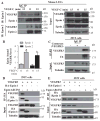
Fig. 6. Epsin interacted with activated VEGFR3 through its UIM domain
A) Mouse LECs were stimulated with VEGF-C for indicated time points and lysed followed by immunoprecipitation with epsin 1 antibodies or control IgG. Interactions were detected by Western blotting with the indicated antibodies. B) Quantification of epsin 1-bound VEGFR3 (n=3 independent experiments) and epsin 2-bound VEGFR3 (n=3 independent experiments). All values are mean ± SD. *p<0.05; One-way ANOVA. C) Lysates from HEK 293T cells expressing VEGFR3 or empty vector were stimulated with VEGF-C and then immunoprecipitated with VEGFR3 antibodies then Western blotted with ubiquitin antibodies (n=3 independent experiments). Ub: ubiquitin. D) Lysates from HEK 293T cells expressing combinations of VEGFR3, HA-epsin 1, HA-epsin 1ΔUIM, or empty vector and stimulated with VEGF-C for 15 minutes were immunoprecipitated with HA antibodies or control IgG and Western blotted with the indicated antibodies (n=3 independent experiments). E) Lysates from HEK 293T cells expressing combinations of VEGFR3, HA-epsin 1, HA-epsin 1ΔUIM, or empty vector and stimulated with VEGF-C were immunoprecipitated with VEGFR3 antibodies or control IgG and Western blotted with the indicated antibodies (n=3 independent experiments).
Fig. 7
Fig. 7. Improved lymphatic valve formation by reducing VEGFR3 abundance or activity in epsin-deficient mice
A–C) Representative whole mount immunostaining of mesenteric lymphatic valves of P6 wild-type mice (A), LEC-iDKO mice (B), and LEC-iDKO-Flt4eGFP/+ mice (C). White arrow indicates potential lymphatic valve region. Scale bar, 50 μm. D) Representative image of lymphatic drainage after FITC-Dextran injection into the footpads of P6 LEC-iDKO-Flt4eGFP/+ mice. Scale bar, 50 μm. E) Quantification of percentage of functional collecting lymphatic vessels assessed as in (C) in P6 wild-type, LEC-iDKO or LEC-iDKO-Flt4eGFP/+ pups (n=3 pups per group analyzed in 3 independent experiments). All values are mean ± SD. *p<0.05; Logistic regression. F–G) Representative whole mount immunostaining of mesenteric lymphatic valves of P6 LEC-DKO mice treated with DMSO (F) or LEC-DKO mice treated with MAZ51 (G). White arrow indicates potential lymphatic valve region. Scale bar, 50 μm. H) Quantification of percentage of V-shape mesenteric lymphatic valves in (A–C) and (F–G) (n=3 pups per group analyzed in 3 independent experiments). All values are mean ± SD. **p<0.01; Logistic regression. I) Representative image of lymphatic drainage after FITC-Dextran injection into footpads of P6 LEC-DKO mice treated with MAZ51. Scale bar, 50 μm. J) Quantification of percentage of functional collecting lymphatic vessels in P6 LEC-DKO pups assessed as in (I) after treatment with DMSO or MAZ51 (n=6 pups per group analyzed in 3 independent experiments). All values are mean ± SD. *p<0.05; Logistic regression. K) Quantification of mean lymphatic vessel diameters in P6 DMSO-treated LEC-DKO, MAZ51-treated LEC-DKO, LEC-iDKO, LEC-iDKO-Flt4eGFP/+ and wild-type intestinal capillary. Statistically significant differences are indicated. All values are mean ± SD (n=3 embryos per group analyzed in 3 independent experiments). *p<0.05; One-way ANOVA.

References

    1. Karpanen T, Alitalo K. Molecular biology and pathology of lymphangiogenesis. Annu Rev Pathol. 2008;3:367–97. - PubMed
    1. François M, Caprini A, Hosking B, Orsenigo F, Wilhelm D, Browne C, Paavonen K, Karnezis T, Shayan R, Downes M, Davidson T, Tutt D, Cheah KS, Stacker SA, Muscat GE, Achen MG, Dejana E, Koopman P. Sox18 induces development of the lymphatic vasculature in mice. Nature. 2008;456:643–7. - PubMed
    1. Lin FJ, Chen X, Qin J, Hong YK, Tsai MJ, Tsai SY. Direct transcriptional regulation of neuropilin-2 by COUP-TFII modulates multiple steps in murine lymphatic vessel development. J Clin Invest. 2010;120:1694–707. - PMC - PubMed
    1. Wigle JT, Oliver G. Prox1 function is required for the development of the murine lymphatic system. Cell. 1999;98:769–78. - PubMed
    1. Srinivasan RS, Oliver G. Prox1 dosage controls the number of lymphatic endothelial cell progenitors and the formation of the lymphovenous valves. Genes Dev. 2011;25:2187–97. - PMC - PubMed

Publication types

MeSH terms