. 2014 Dec;5(6):743-51.
doi: 10.1007/s13244-014-0364-y. Epub 2014 Oct 15.

Vocal cord paralysis: anatomy, imaging and pathology

Affiliations

Vocal cord paralysis: anatomy, imaging and pathology

J W Dankbaar et al. Insights Imaging. 2014 Dec.

Abstract

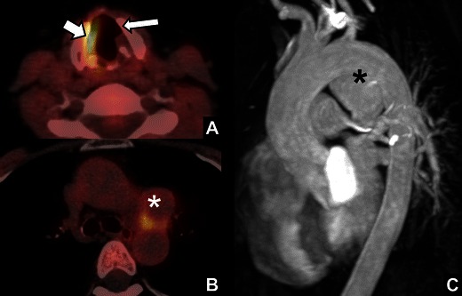
Vocal cord paralysis (VCP) can be caused by any process that interferes with the normal function of the vagal nerves or recurrent laryngeal nerves. It may be a first sign of extensive and severe pathology. Radiologists must therefore be able to recognise the imaging findings of VCP and know the course of the vagal and recurrent laryngeal nerves. This review focuses on the anatomy and imaging evaluation of these nerves and thereby the possible sites for pathology causing VCP. The imaging characteristics and imaging mimics of VCP are discussed and cases from daily practice illustrating causes of VCP are presented.

Teaching points: • Vocal cord paralysis may be the first presentation of severe pathology. • Radiologists must be aware of imaging characteristics and mimics of vocal cord paralysis. • Lesions along the vagal nerves and recurrent laryngeal nerves can cause vocal cord paralysis.


Figures

Fig. 1
Fig. 1
Normal appearance of the vocal cords. a Axial CT images during quiet breathing showing the anterior commissure (short arrow), the posterior commissure (long arrow) and the true vocal cords (T). b Image at the same level during breath-hold. c Normal appearance of the laryngeal ventricles on coronal reformat: right laryngeal ventricle (arrow); left true vocal cord (T); left false vocal cord (F)
Fig. 2
Fig. 2
The vagal nerves in the posterior fossa.Axial heavily T2-weighted thin-slice MR image through the brainstem with the position of the nucleus ambiguus (white circles), the exit of the vagal nerves from the olivary sulcus (curved arrow), the cisternal segment of the vagal nerves (long arrow), and the pars nervosa of the jugular foramen (short arrow)
Fig. 3
Fig. 3
The course of the vagal nerves and recurrent laryngeal nerves. a Coronal MIP reformat showing the expected course of the vagal nerves bilaterally within the carotid sheath and proximal parts of the recurrent laryngeal nerves (dotted lines). The right recurrent laryngeal nerve branches just caudally to the right subclavian artery (short arrow) and crosses the right subclavian artery towards the tracheo-oesophageal groove. The left recurrent laryngeal nerve (long arrow) runs below the aortic arch. b Contrast enhanced CT of the skull base showing the jugular foramen with the jugular vein (long arrow), the pars nervosa (short arrow) and the internal carotid artery (*). c The carotid sheath with the internal carotid artery (A), the jugular vein (V) and the expected position of vagal nerve (*). d At the level of the subclavian arteries (S) the vagal nerves (long arrows) lie anteriorly in the upper mediastinum. At this level, the recurrent laryngeal nerves run more posteriorly in the tracheo-oesophageal groove (short arrows). e The left recurrent laryngeal nerve crosses the aortic arch from anterior to posterior (curved arrow) at the level of the aorto-pulmonary window (AP)
Fig. 4
Fig. 4
Specific imaging characteristics of vocal cord paralysis (right side). a Widening of the right laryngeal ventricle (arrow). b Medial deviation and thickening of the right aryepiglottic fold (arrow). c Dilatation of the right piriform sinus (arrow)
Fig. 5
Fig. 5
Supportive imaging characteristics of vocal cord paralysis (VCP). a Pointed right vocal cord (long arrow) on coronal reformat due to atrophy; note the widening of the ipsilateral laryngeal ventricle (short arrow). b Subglottic fullness due to sagging of the paralysed right vocal cord (*). c Paramedian position of the paralysed right vocal cord (*)
Fig. 6
Fig. 6
Mimics of VCP. a Tilted axial reformat through the larynx showing subglottic air (arrow) projecting anterior to the angulated normal right true vocal cord, mimicking a widened laryngeal ventricle seen in VCP. b Small squamous cell carcinoma of the left vocal cord (*) mimicking the paramedian position seen in VCP
Fig. 7
Fig. 7
FDG-PET findings in VCP. a Non-contrast CT image showing medial deviation and thickening of the left aryepiglottic fold (*) indicative of left VCP. b Fused PET-CT showing FDG uptake in the right vocal cord (arrow) due to compensatory hypertrophy. This should not be confused with disease. Note the paramedian position of the left true vocal cord (T)
Fig. 8
Fig. 8
Bilateral VCP. a Axial CT reformat showing bilateral medial deviation and thickening of the aryepiglottic fold (arrows) and bilateral dilatation of the piriform sinuses (*). b Coronal CT reformat showing bilateral widening of the laryngeal ventricles (arrows)
Fig. 9
Fig. 9
Brainstem infarction.A 55-year-old woman presenting with VCP after endovascular coiling of a right vertebral artery aneurysm. DWI at the level of the medulla oblongata showed a small area of restricted diffusion directly at the site of the right vagal nerve nucleus, indicative of infarction (arrow)
Fig. 10
Fig. 10
Skull base metastasis.A 66-year-old man with a history of oropharynx carcinoma treated with radiation therapy 1 year earlier presenting with left VCP. FDG-PET revealed a hypermetabolic lesion in the left skull base presumed to be a metastasis. a Axial T1-weighted contrast-enhanced images at the level of the skull base showed a heterogeneously enhancing mass in the left cerebellomedullary cistern (arrow). The mass shows infiltrative growth into the skull base and jugular foramen, and thereby the vagal nerve, causing left VCP. The imaging findings were suspicious for metastatic disease. b Follow-up T1-weighted contrast-enhanced images with fat saturation showed progression of the skull-base mass with infiltrative growth into the nasopharynx (long arrow) and mass effect on the cerebellum and pons (short arrow)
Fig. 11
Fig. 11
Osteomyelitis of the skull base. A 74-year-old man presenting with slowly progressive symptoms of left hypoglossal nerve palsy and left VCP. CT and MR images at the level of the skull base (not displayed) showed fluid in the mastoid and middle ear bilaterally. a Contrast-enhanced CT and b) T1-weighted contrast-enhanced images with fat saturation showed infiltration of the area between the petrous apex and the temporo-mandibular joint by a destructive enhancing mass (*). The imaging findings suggested osteomyelitis and soft tissue infection. The jugular foramen and thereby the vagal nerve were obviously involved, causing VCP. The patient underwent left-sided exploration and mastoidectomy revealing massive infection of the middle ear, skull-base and neck
Fig. 12
Fig. 12
Glomus jugulare tumour.A 42-year-old man presenting with slowly progressive symptoms of left VCP with no other apparent laryngeal pathology. a Coronal T2-weighted MRI images and b T1-weighted contrast-enhanced fat-saturated images showed a T2 hyperintense avidly enhancing, smooth lobulated mass, with a salt and pepper appearance at the left jugular foramen (arrows) typical of glomus jugulare tumour
Fig. 13
Fig. 13
Lymph node metastasis in the neck from oropharyngeal squamous cell carcinoma.A 66-year-old man with a history of smoking and alcohol abuse presenting with a swelling on the right side of the neck and right VCP. Coronal contrast enhanced CT images showed a large lymph node conglomerate at level IIb (arrow) on the right side of the neck, with necrosis, extra nodal spread and extensive involvement of the right carotid space explaining the right VCP in this patient
Fig. 14
Fig. 14
Paratracheal lymph node metastasis from oesophageal carcinoma. A 53-year-old man with a history of heavy smoking and drinking presenting with left VCP. a Coronal contrast-enhanced CT images showed a mass in the left TE groove in the upper mediastinum in the course of the left recurrent laryngeal nerve (arrow). b Sagittal fused FDG-PET-CT images showed avid FDG uptake in the mass (short arrow) and in a thickened portion of oesophagus just below the carina (long arrow). The patient was diagnosed with oesophageal carcinoma with left paratracheal lymph node metastasis causing left sided VCP
Fig. 15
Fig. 15
Lung cancer with involvement of the aorto-pulmonary (AP) window.72 year old male smoker with a history of squamous cell carcinoma of the left ear presenting with functional dysphonia caused by left VCP. a Axial contrast-enhanced CT of the larynx showing complete atrophy of the left true vocal cord (short arrow) and hypertrophy of the right true vocal cord (*). b Images at the level of the AP window showed a large supra-hilar mass in the left lung infiltrating the AP-window (arrow), thereby explaining left VCP in this patient. Biopsy revealed a non-small cell lung carcinoma
Fig. 16
Fig. 16
Aortic arch aneurysm. A 44-year-old patient with a history of systemic lupus erythematosus and herpes zoster infection presenting with symptoms of left VCP. Because of the patient’s history, a FDG-PET with low-dose CT was obtained. a Fused PET-CT image at the level of the cricoid showed widening of the left laryngeal ventricle (long arrow), note the increased FDG-uptake in the unaffected right vocal cord due to hypertrophy (short arrow). b Images at the level of the aortic arch showed a mass in the AP-window without FDG uptake (*). c Contrast-enhanced MR angiography of the aortic arch showed a saccular aneurysm (*) causing compression of the vagal nerve at the site of the branching of the left laryngeal recurrent nerve, thereby causing VCP

References

    1. Rosenthal LH, Benninger MS et al (2007) Vocal fold immobility: a longitudinal analysis of etiology over 20 years. Laryngoscope 117:1864–1870 - PubMed
    1. Richardson BE, Bastian RW (2004) Clinical evaluation of vocal fold paralysis. Otolaryngol Clin North Am 37:45–58 - PubMed
    1. Myssiorek D (2004) Recurrent laryngeal nerve paralysis: anatomy and etiology. Otolaryngol Clin North Am 37:25–44 - PubMed
    1. Collazo-Clavell ML, Gharib H, et al. Relationship between vocal cord paralysis and benign thyroid disease. Head Neck. 1995;17:24–30. doi: 10.1002/hed.2880170106. - DOI - PubMed
    1. ten Donkelaar HJ (2007) Klinische Anatomie en Embryologie. Reed Business, Amsterdam. ISBN: 9789035229013

LinkOut - more resources