. 2015 Mar 1;24(5):550-64.
doi: 10.1089/scd.2014.0310. Epub 2014 Dec 2.

Epigenetic disruptions of histone signatures for the trophectoderm and inner cell mass in mouse parthenogenetic embryos

Affiliations

Epigenetic disruptions of histone signatures for the trophectoderm and inner cell mass in mouse parthenogenetic embryos

Yi-Hui Chen et al. Stem Cells Dev. .

Abstract

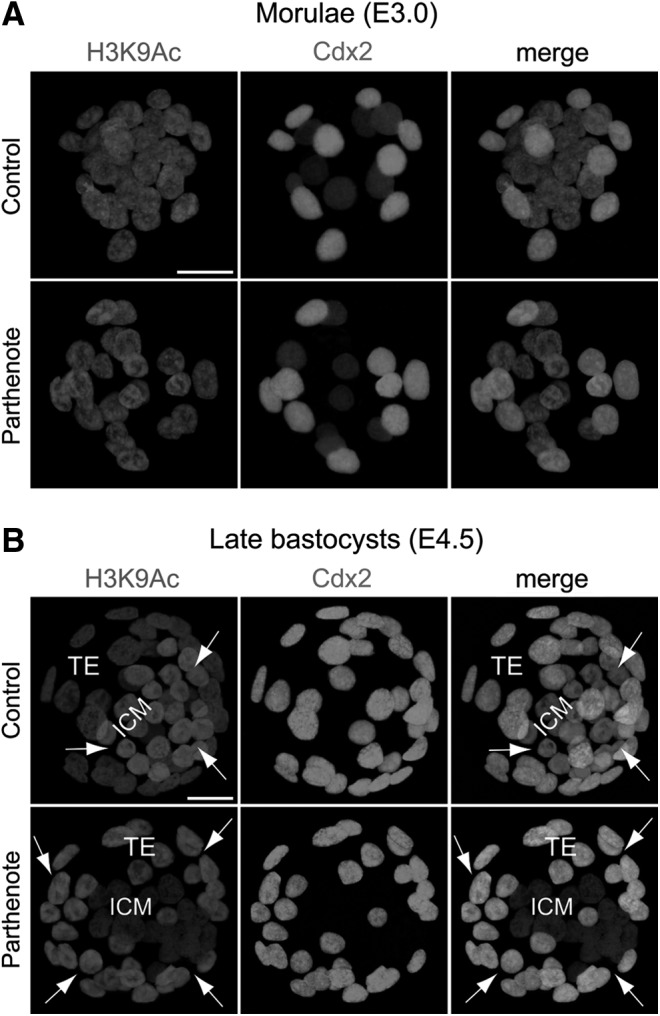
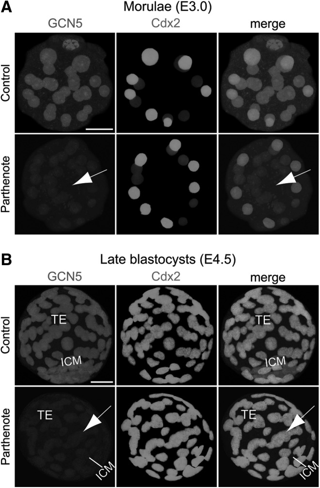
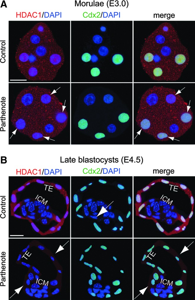
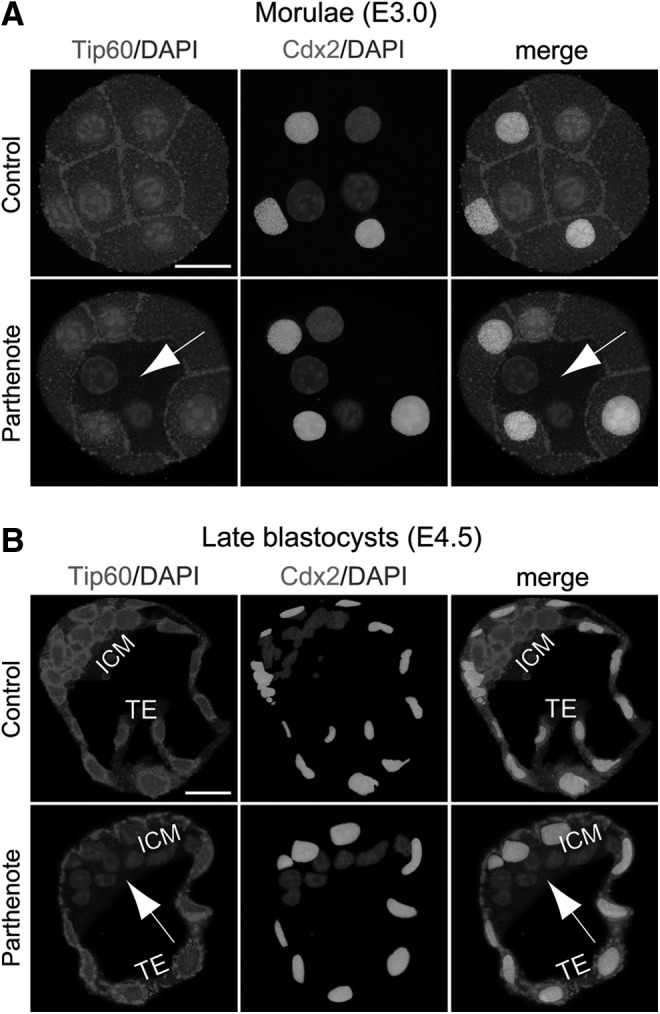
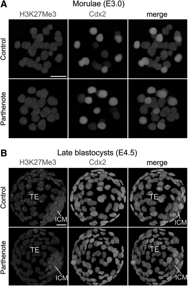
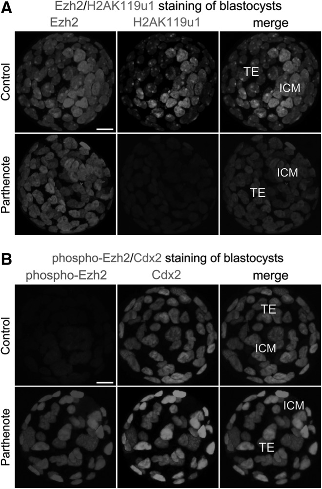
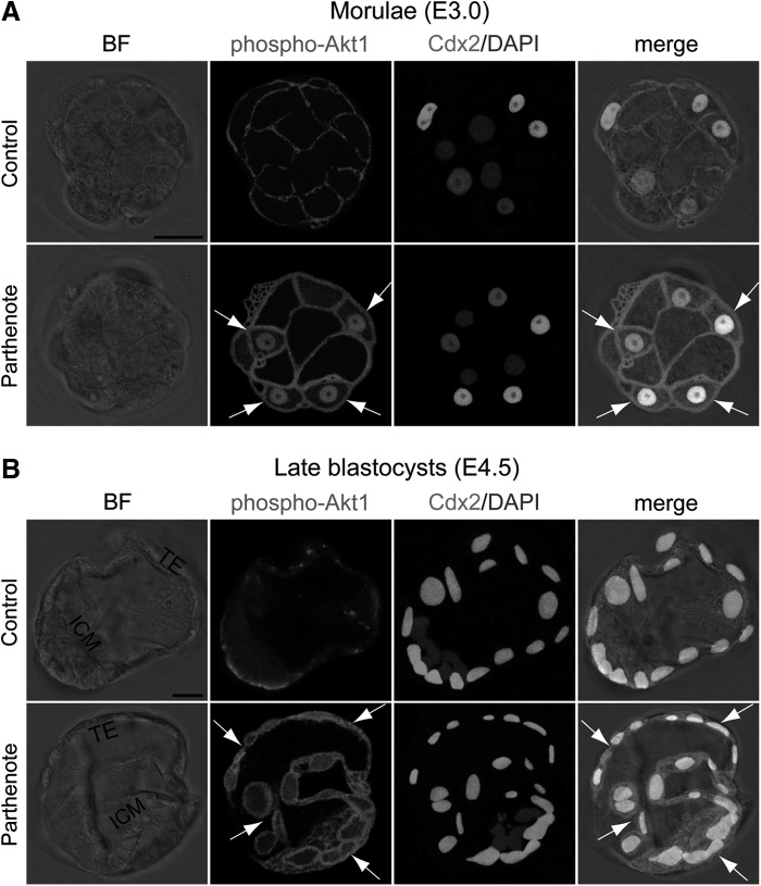
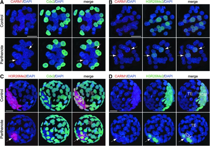
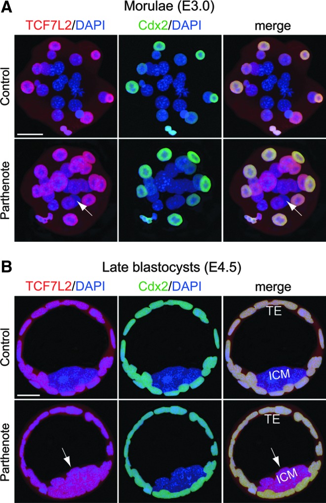
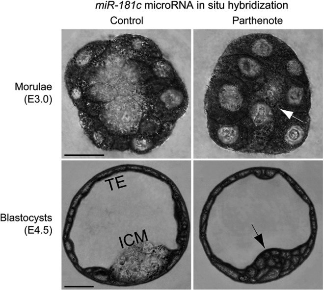
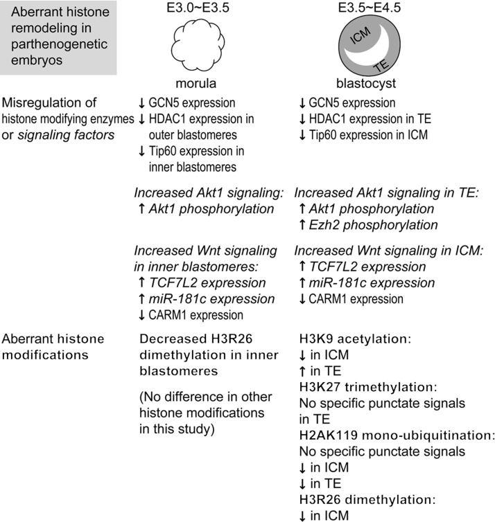
Epigenetic asymmetry has been shown to be associated with the first lineage allocation event in preimplantation development, that is, the formation of the trophectoderm (TE) and inner cell mass (ICM) lineages in the blastocyst. Since parthenogenesis causes aberrant segregation between the TE and ICM lineages, we examined several development-associated histone modifications in parthenotes, including those involved in (i) transcriptional activation [acetylated histone H3 lysine 9 (H3K9Ac) and lysine 14 (H3K14Ac), trimethylated histone H3 lysine 4 (H3K4Me3), and dimethylated histone H3 arginine 26 (H3R26Me2)] and (ii) transcriptional repression [trimethylated histone H3 lysine 9 (H3K9Me3) and lysine 27 (H3K27Me3), and mono-ubiquitinated histone H2A lysine 119 (H2AK119u1)]. Here, we report that in parthenotes, H3R26Me2 expression decreased from the morula stage, while expression patterns and levels of H3K9Ac, H3K27Me3, and H2AK119u1 were unchanged until the blastocyst stage; whereas H3K14Ac, H3K4Me3, and H3K9Me3 showed normal patterns and levels of expressions. Relative to the decrease of H3K9Ac in the ICM and increase in the TE of parthenotes, we detected reduced expression of TAT-interactive protein 60 acetyltransferase and histone deacetylase 1 deacetylase in the ICM and TE of parthenotes, respectively. Relative to the decrease of H3R26Me2, we also observed decreased expression of coactivator-associated arginine methyltransferase 1 methyltransferase and increased expression of the Wnt effector transcription factor 7L2 and miR-181c microRNA in parthenotes. Furthermore, relative to the decrease in H3K27Me3 and H2AK119u1, we found increased phosphorylation of Akt1 and enhancer of zeste homolog 2 in parthenogenetic TE. Therefore, our findings that histone signatures are impaired in parthenotes provide a mechanistic explanation for aberrant lineage segregation and TE defects.


Figures

<b>FIG. 1.</b>
FIG. 1.
Reversed asymmetrical distribution of acetylated histone H3 lysine 9 (H3K9Ac) in parthenogenetic blastocysts. Double immunostaining of acetylated histone H3K9 and Cdx2 revealed relatively homogeneous staining among all nuclei of both control (normal fertilized) and parthenogenetic morulae (E3.0) (A) and asymmetrical staining between the inner cell mass (ICM) and trophectoderm (TE) of control and parthenogenetic blastocysts (E4.5) (B). Cdx2 was detected in the outer nuclei of morulae and the TE of blastocysts. Note that the asymmetrical distribution of H3K9Ac between the ICM and the TE was reversed in parthenogenetic blastocysts as compared with control blastocysts (B); that is, the H3K9Ac level was apparently higher in the ICM than TE in controls, but was significantly lower in the ICM than TE in parthenotes. Arrows indicate cells with a relatively high H3K9Ac level. Scale bars: 20 μm.
<b>FIG. 2.</b>
FIG. 2.
Decreased expression of GCN5 acetyltransferase in parthenogenetic morulae and blastocysts. Double immunostaining against GCN5 and Cdx2 revealed significantly decreased GCN5 expression (indicated by arrows) throughout the whole parthenogenetic embryos (including both the outer Cdx2+ and inner Cdx2- blastomeres) compared with control embryos at both the morula (A) and blastocyst (B) stages. Scale bars: 20 μm.
<b>FIG. 3.</b>
FIG. 3.
Decreased expression of histone deacetylase 1 (HDAC1) deacetylase in the outer blastomeres and TE of parthenogenetic embryos. Double immunostaining for HDAC1 (red) and Cdx2 (green) indicated that HDAC1 was expressed at a significantly higher level in the Cdx2+ blastomeres (ie, the outer blastomeres of morulae and TE of blastocysts) (A) than in the Cdx2- blastomeres (ie, the inner blastomeres of morulae and ICM of blastocysts) (B) in control embryos. At the morula stage, HDAC1 expression was detectable in both the nuclei and cytoplasm of parthenogenetic outer blastomeres, although at a reduced level compared with control blastomeres [indicated by arrows in (A)]. At the blastocyst stage, the reduction of HDAC1 expression in parthenotes was magnified so that both the TE and ICM displayed only a background staining level of HDAC1 in the cytoplasm [indicated by arrows in (B)]. Scale bars: 20 μm.
<b>FIG. 4.</b>
FIG. 4.
Decreased expression of TAT-interactive protein 60 (Tip60) acetyltransferase in the inner blastomeres and ICM of parthenogenetic embryos. Double immunostaining against Tip60 and Cdx2 indicated that, in parthenogenetic embryos, Tip60 expression was dramatically reduced in the Cdx2 blastomeres (indicated by arrows), including the inner blastomeres of morulae (A) and ICM of blastocysts (B), compared with control embryos, which exhibited strong and homogeneous Tip60 expression throughout the whole embryos. Scale bars: 20 μm.
<b>FIG. 5.</b>
FIG. 5.
Absence of punctate staining against trimethylated histone H3 lysine 27 (H3K27Me3) in nuclei of the parthenogenetic TE. Immunostaining against dimethylated histone H3K27 revealed multiple punctate signals in all nuclei of both control (normally fertilized) and parthenogenetic morulae (E3.0) (A). At the blastocyst stage, however, the single punctate staining pattern of H3K27Me3 observed in all nuclei of the control TE (expressing Cdx2 with fluorescence) was absent in the parthenogenetic TE (B). Note the higher staining level in the ICM as compared with the TE in both control and parthenogenetic blastocysts (B). Scale bars: 20 μm.
<b>FIG. 6.</b>
FIG. 6.
Dramatically decreased mono-ubiquitination of histone H2A lysine 119 (H2AK119u1) and phosphorylation of enhancer of zeste homolog 2 (Ezh2) methyltransferase in parthenogenetic blastocysts. Immunostaining was used to reveal that levels of total Ezh2 methyltransferase were similar between the nuclei of the TE of control and parthenogenetic blastocysts (A). The level of ubiquitinated histone H2AK119 was significantly decreased in both the TE and ICM of parthenogenetic blastocysts (A), while the level of serine 21-phosphorylated Ezh2 was dramatically higher in the parthenogenetic TE than in the control TE (B). Note that the ICM in both control and parthenogenetic blastocysts exhibited only a background level of Ezh2 phosphorylation (B). Scale bars: 20 μm.
<b>FIG. 7.</b>
FIG. 7.
Increased phosphorylation of Akt1 in parthenogenetic morulae and blastocysts. Double immunostaining against serine 473-phosphorylated Akt1 and Cdx2 revealed that the phosphorylation level of Akt1 was greatly increased at the plasma membrane and in the cytoplasm of parthenogenetic Cdx2+ blastomeres, during both the morula (E3.0) (A) and blastocyst (E4.5) (B) stages (indicated by arrows). While Ser473-phospho-Akt1 staining was present in the nuclei of most outer blastomeres in parthenogenetic morulae [nuclei indicated by arrows in (A)], nuclear staining of phospho-Akt1 was absent from parthenogenetic blastocysts [arrows in (B)]. Scale bars: 20 μm.
<b>FIG. 8.</b>
FIG. 8.
Decreased coactivator-associated arginine methyltransferase 1 (CARM1) expression and dimethylation of histone H3 arginine 26 (H3R26Me2) in parthenogenetic embryos. (A, B) Immunostaining against CARM1 (red) revealed greatly decreased numbers of positive cells in parthenogenetic morulae [indicated by arrows in (A) and (B)]. Immunostaining signals against dimethylated histone H3R26 [green in (B)] revealed co-localization with those against CARM1 [red in (B)], and a dramatic decrease in the number of positive cells in the parthenogenetic morula [indicated by arrows in (B)]. (C, D) Immunostaining against dimethylated histone H3R26 [red in (C) and green in (D)] revealed significantly lower signals in parthenogenetic blastocysts, primarily in the ICM (indicated by arrows). Immunostaining signals against CARM1 methyltransferase [red in (D)] were co-localized with those of H3R26Me2 [green in (B)], and were also significantly decreased in parthenogenetic blastocysts [this decrease was also primarily in the ICM, as indicated by the arrows in (D)]. Scale bars: 20 μm.
<b>FIG. 9.</b>
FIG. 9.
Increased expression of the Wnt effector transcription factor 7L2 (TCF7L2) in parthenogenetic morulae and blastocysts. Double immunostaining against TCF7L2 (red) and Cdx2 (green) revealed dramatically increased TCF7L2 expression (indicated by arrows) in the inner blastomeres of parthenogenetic morulae (A) and parthenote ICM (B) as compared with control embryos. On the other hand, TCF7L2 expression in the outer blastomeres of morulae and TE was comparable between control and parthenogenetic embryos (A, B). Scale bars: 20 μm.
<b>FIG. 10.</b>
FIG. 10.
Increased expression of the CARM1 inhibitor miR-181c microRNA in parthenogenetic morulae and blastocysts. RNA in situ hybridization was performed using an LNA-modified DNA oligonucleotide probe against miR-181c microRNA. Expression of miR-181c was dramatically increased in the inner blastomeres of parthenogenetic morulae and the ICM of parthenogenetic blastocysts (indicated by arrows), as compared with control embryos. On the other hand, miR-181c expression in the outer blastomeres of morulae and TE was comparable between control and parthenogenetic embryos. Scale bars: 20 μm.
<b>FIG. 11.</b>
FIG. 11.
Summary of aberrant histone modifications and misregulation of histone-modifying enzymes and associated signaling factors in parthenogenetic morulae and blastocysts. Of the histone modifications analyzed in this study, only the dimethylation level of histone H3 arginine 26 (H3R26) was different between control and parthenogenetic embryos at the morula stage. At the blastocyst stage, however, levels of the following histone modifications were altered in parthenotes: H3R26 dimethylation, histone H3 lysine 9 (H3K9) acetylation, histone H3 lysine 27 (H3K27) trimethylation, and histone H2A lysine 119 (H2AK119) mono-ubiquitination. Decreased dimethylation of H3R26 in parthenotes may be due to decreased expression of CARM1 methyltransferase, which may result from the observed increase in TCF7L2 (a component of Wnt signaling) and expression of miR-181c microRNA at both the morula and blastocyst stages. Decreased trimethylation of H3K27 in parthenote TE, on the other hand, is associated with decreased mono-ubiquitination of H2AK119 and increased phosphorylation of Ezh2 methyltransferase, which may result from the observed increase in phosphorylation of Akt1 in parthenogenetic blastocysts. In addition, we found that H3K9 acetylation is decreased in parthenote ICM and increased in parthenote TE as compared with control blastocysts, whereas the underlying molecular mechanism remains to be elucidated.

Similar articles

Cited by

References

    1. Cassar G, John TM. and Etches RJ. (1998). Observations on ploidy of cells and on reproductive performance in parthenogenetic turkeys. Poult Sci 77:1457–1462 - PubMed
    1. Fujita MK. and Moritz C. (2009). Origin and evolution of parthenogenetic genomes in lizards: current state and future directions. Cytogenet Genome Res 127:261–272 - PubMed
    1. Parker HM, Kiess AS, Wells JB, Young KM, Rowe D. and McDaniel CD. (2010). Genetic selection increases parthenogenesis in Chinese painted quail (Coturnix chinensis). Poult Sci 89:1468–1472 - PubMed
    1. Sinclair EA, Pramuk JB, Bezy RL, Crandall KA. and Sites JW, Jr., (2010). DNA evidence for nonhybrid origins of parthenogenesis in natural populations of vertebrates. Evolution 64:1346–1357 - PubMed
    1. Cheng L. (2008). More new lines of human parthenogenetic embryonic stem cells. Cell Res 18:215–217 - PubMed

Publication types

MeSH terms

Substances

LinkOut - more resources