Review
. 2015 Mar;1338(1):94-114.
doi: 10.1111/nyas.12553. Epub 2014 Oct 14.

Autoimmune encephalopathies

Affiliations
Review

Autoimmune encephalopathies

Frank Leypoldt et al. Ann N Y Acad Sci. 2015 Mar.

Abstract

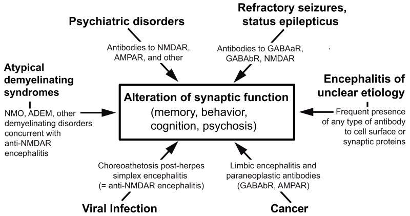
Over the past 10 years, the continual discovery of novel forms of encephalitis associated with antibodies to cell-surface or synaptic proteins has changed the paradigms for diagnosing and treating disorders that were previously unknown or mischaracterized. We review here the process of discovery, the symptoms, and the target antigens of 11 autoimmune encephalitic disorders, grouped by syndromes and approached from a clinical perspective. Anti-N-methyl-d-aspartate receptor (NMDAR) encephalitis, several subtypes of limbic encephalitis, stiff-person spectrum disorders, and other autoimmune encephalitides that result in psychosis, seizures, or abnormal movements are described in detail. We include a novel encephalopathy with prominent sleep dysfunction that provides an intriguing link between chronic neurodegeneration and cell-surface autoimmunity (IgLON5). Some of the caveats of limited serum testing are outlined. In addition, we review the underlying cellular and synaptic mechanisms that for some disorders confirm the antibody pathogenicity. The multidisciplinary impact of autoimmune encephalitis has been expanded recently by the discovery that herpes simplex encephalitis is a robust trigger of synaptic autoimmunity, and that some patients may develop overlapping syndromes, including anti-NMDAR encephalitis and neuromyelitis optica or other demyelinating diseases.

Keywords: anti-NMDAR antibodies; autoimmune encephalitis; limbic encephalitis; psychosis; treatment.


Figures

Figure 1
Figure 1
Process of discovery, antigen immunoprecipitation, and development of a diagnostic test. Dissociated rat hippocampal neurons maintained in vitro and incubated (live, non-permeabilized) with CSF of a patient. Note the intense reactivity of patient’s antibodies with cell surface antigens (A); scale bar = 10 μm. Confocal microscopy suggests that the antigens are concentrated in clusters along dendrites (B); scale bar = 5 μm. Precipitation of these antigens using two patients’ CSF antibodies is shown in a gel in which proteins are visualized with EZBlue (C). Note that patients’ antibodies (P1, P2) precipitated a single band at ~100 kDa; this band is not seen in the precipitate of a control individual (N). Analysis of the 100 kDa band by mass spectrometry demonstrated sequences derived from GluR1/GluR2 subunits of the AMPAR. The ~50 kDa band across all samples corresponds to IgG. Transfer of the proteins to nitro-cellulose and Western blot with GluR1 and GluR2 antibodies confirmed that the 100 kDa band contained both GluR1 and GluR2 subunits (panels in D). Further validation of the antigen was done in heterologous cells expressing GluR1/2, showing reactivity with patient’s antibodies (green), a monoclonal antibody (red), and the merged reactivities (yellow). Adapted from Lai and colleagues with permission.
Figure 2
Figure 2
Comparison of MRIs of patients with limbic encephalitis and different cell surface autoantibodies with the MRI of a patient with multifocal encephalitis and GABAaR receptor antibodies. The MRIs of the three cases with limbic encephalitis correspond to a patient with LGI1 antibodies (A), a patient with AMPAR antibodies (B), and a patient with GABAb-R antibodies (C). In contrast, note that the MRI of the patient with GABAaR antibodies shows multifocal cortical-subcortical FLAIR abnormalities (D). Panels A–C, from Lancaster and colleagues, with permission; panel D from Petit-Pedrol and colleagues with permission.
Figure 3
Figure 3
Sequence of syndrome development in patients with anti-NMDAR encephalitis. During the first month of the disease most patients with anti-NMDAR encephalitis progressively develop the sequence of symptoms shown in the graph. Adapted from Dalmau and colleagues.
Figure 4
Figure 4
Synaptic and behavioral effects of antibodies from patients with anti-NMDAR encephalitis. (A–C) AMPA and NMDA receptors are localized in the postsynaptic membrane and are clustered at the postsynaptic density A. Patient antibodies in the CNS bind selectively to NMDA receptors at the synapse as well as extrasynaptic receptors. This binding leads to receptor cross-linking B. NMDA receptors that have been bound and cross-linked by antibodies are internalized, resulting in a decrease of surface, synaptically localized NMDA receptors. Other synaptic components, such as postsynaptic AMPA receptor clusters, PSD-95, as well as presynaptic terminals, dendrite branches, dendritic spines and cell viability, are unaffected C. Thus patient anti-NMDA receptor antibodies lead to a rapid, selective excision of NMDA receptors from neuronal membranes. This effect is titer-dependent and reverses after antibody titers are reduced (not shown). (D–E) Rodent cultured neurons treated with control CSF or patient’s CSF for 3 days, and subsequently stained for postsynaptic GluN1 to label NMDA receptor clusters, VGlut to label presynaptic sites, and PSD-95 as a postsynaptic marker. Note that patient’s CSF cause a decrease in dendritic GluN1 cluster density, while VGlut and PSD-95 cluster density remain unchanged. (F–G) Whole-cell patch recordings of miniature excitatory postsynaptic currents (mEPSCs) that consist of a fast AMPA receptor–mediated component and a slower later component mediated by NMDA receptors and is APV sensitive. Compared with neurons incubated with CFS control F, those treated with CSF from a patient with NMDA receptor antibodies show a loss of the APV-sensitive NMDA receptor component E. H: Progressive memory deficit assessed by standard object recognition test, in a group of 5 mice treated with cerebroventricular infusion of CSF from patients with anti-NMDAR encephalitis (grey circles) compared with a group of 5 mice treated with CSF from control subjects without NMDAR antibodies (white circles). Antibodies were continuously infused for 14 days; the peak of memory deficit was on day 18, followed by progressive clinical recovery. Panels (A–G) obtained from Moscato and colleagues, with permission. Panel H provide by Planaguma and colleagues (unpublished data).
Figure 5
Figure 5
Interaction of LGI1 with synaptic proteins. Secreted LGI1 interacts at the presynapse with ADAM23 and at the postsynapse with ADAM22. The figure is based on studies indicating that LGI1 co-precipitates with other proteins including the presynaptic Kv1 potassium channels and a variety of pre- and post-synaptic scaffolding proteins. Adapted from Lancaster and colleagues, with permision.
Figure 6
Figure 6
Associations, trigers, and common symptoms of autoimmune encephalitis: impact on multiple disciplines
Figure 7
Figure 7
Plasma cell infiltrates in the brain of a patient with anti-NMDAR encephalitis. Paraffin embedded sections of brain biopsy of a patient with anti-NMDAR encephalitis. CD138+ cells (plasma cells/plasmablasts) are present in perivascular, Virchow-Robin (A), and interstitial spaces (B). In Virchow-Robin spaces (A) the CD138+ cells are in perivascular regions (arrows) and along the tissue surface (arrow heads) that delineates spaces containing CSF and small vessels (v). The plasma cells/plasmablasts indicated with arrows are amplified in the inset in A. Scales = 20 μm. Adapted from Martinez-Hernandez and colleagues, with permision.

References

    1. Lancaster E, Dalmau J. Neuronal autoantigens-pathogenesis, associated disorders and antibody testing. Nat. Rev. Neurol. 2012;8:380–390. - PMC - PubMed
    1. Moscato EH, Jain A, Peng X, et al. Mechanisms underlying autoimmune synaptic encephalitis leading to disorders of memory, behavior and cognition: insights from molecular, cellular and synaptic studies. Eur. J. Neurosci. 2010;32:298–309. - PMC - PubMed
    1. Panzer JA, Gleichman AJ, Lynch DR. Glutamatergic autoencephalitides: an emerging field. J. Neural. Transm. 2014 In press. - PMC - PubMed
    1. Lindstrom JM, Engel AG, Seybold ME, et al. Pathological mechanisms in experimental autoimmune myasthenia gravis. II. Passive transfer of experimental autoimmune myasthenia gravis in rats with anti-acetylcholine receptor antibodies. J. Exp. Med. 1976;144:739–753. - PMC - PubMed
    1. Titulaer MJ, Lang B, Verschuuren JJ. Lambert-Eaton myasthenic syndrome: from clinical characteristics to therapeutic strategies. Lancet Neurol. 2011;10:1098–1107. - PubMed

Publication types

MeSH terms