. 2014 Aug 27;15(8):428.
doi: 10.1186/s13059-014-0428-9.

Metastatic tumor evolution and organoid modeling implicate TGFBR2 as a cancer driver in diffuse gastric cancer

Metastatic tumor evolution and organoid modeling implicate TGFBR2 as a cancer driver in diffuse gastric cancer

Lincoln D Nadauld et al. Genome Biol. .

Abstract

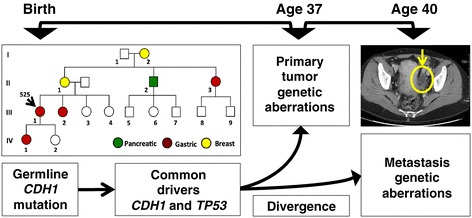
Background: Gastric cancer is the second-leading cause of global cancer deaths, with metastatic disease representing the primary cause of mortality. To identify candidate drivers involved in oncogenesis and tumor evolution, we conduct an extensive genome sequencing analysis of metastatic progression in a diffuse gastric cancer. This involves a comparison between a primary tumor from a hereditary diffuse gastric cancer syndrome proband and its recurrence as an ovarian metastasis.

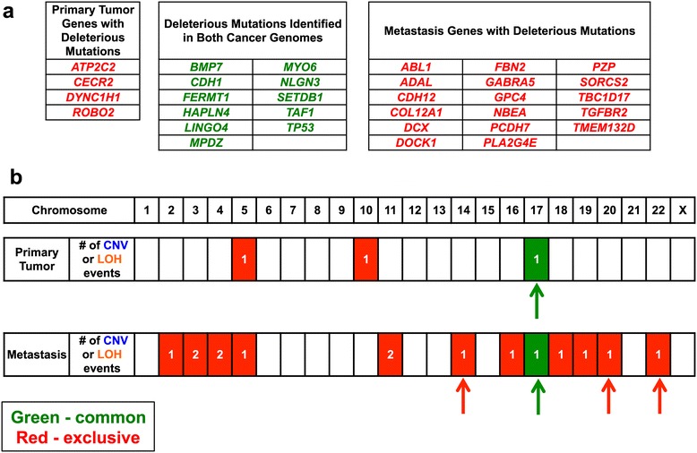
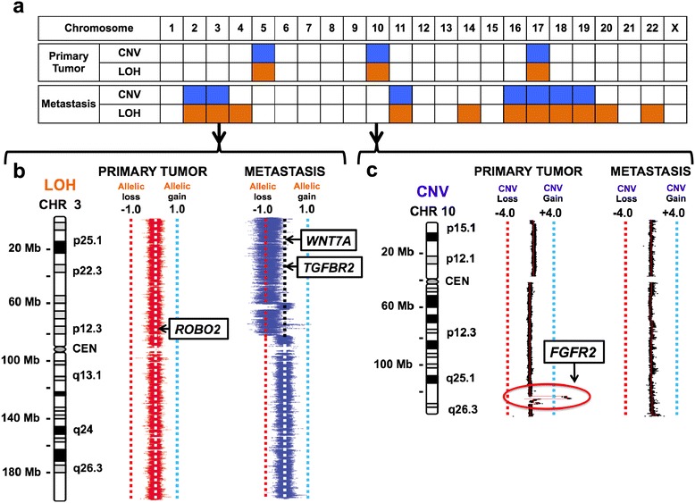
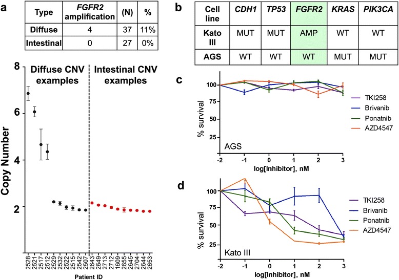
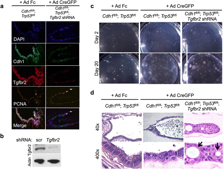
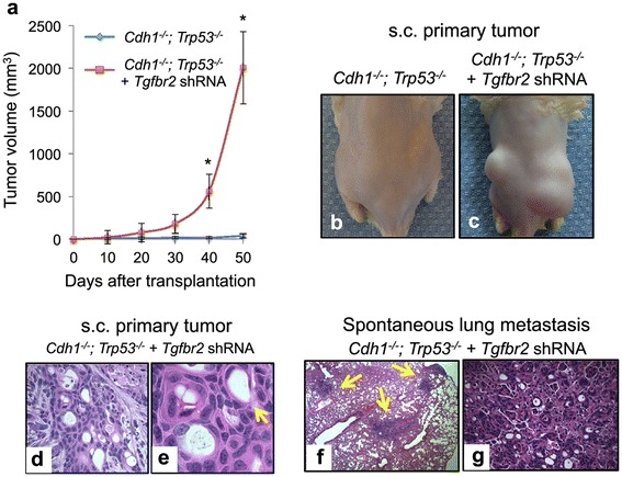
Results: Both the primary tumor and ovarian metastasis have common biallelic loss-of-function of both the CDH1 and TP53 tumor suppressors, indicating a common genetic origin. While the primary tumor exhibits amplification of the Fibroblast growth factor receptor 2 (FGFR2) gene, the metastasis notably lacks FGFR2 amplification but rather possesses unique biallelic alterations of Transforming growth factor-beta receptor 2 (TGFBR2), indicating the divergent in vivo evolution of a TGFBR2-mutant metastatic clonal population in this patient. As TGFBR2 mutations have not previously been functionally validated in gastric cancer, we modeled the metastatic potential of TGFBR2 loss in a murine three-dimensional primary gastric organoid culture. The Tgfbr2 shRNA knockdown within Cdh1-/-; Tp53-/- organoids generates invasion in vitro and robust metastatic tumorigenicity in vivo, confirming Tgfbr2 metastasis suppressor activity.

Conclusions: We document the metastatic differentiation and genetic heterogeneity of diffuse gastric cancer and reveal the potential metastatic role of TGFBR2 loss-of-function. In support of this study, we apply a murine primary organoid culture method capable of recapitulating in vivo metastatic gastric cancer. Overall, we describe an integrated approach to identify and functionally validate putative cancer drivers involved in metastasis.


Figures

Figure 1
Figure 1
Family and clinical history of a Mendelian diffuse gastric cancer. The pedigree of the index patient 525 (III-1) is depicted. Tumor types are indicated by color including green for pancreatic cancer, red for diffuse gastric cancer, and yellow for breast cancer. The patient presented with her primary gastric cancer at the age of 37 years. Three years later she presented with an abdominal discomfort. Contrast-enhanced CT scan of the pelvis identified a left ovary mass (yellow circle) that was confirmed on biopsy to be a diffuse gastric cancer metastasis (that is, Krukenberg tumor). During the course of metastatic tumor evolution, a number of known and candidate cancer driver events delineated the tumor evolution and genetic divergence of the metastasis from the primary tumor.
Figure 2
Figure 2
Comparison of the genetic aberrations in the primary tumor and metastasis. Common versus exclusive genetic aberrations are compared between the two tumor genomes. (a) Genes with coding mutations having a potential deleterious impact are listed. These genes are classified based on whether they are exclusive (red characters) or common (green characters) to the primary tumor and metastasis. The mutations all lead to changes in the amino acid composition of the gene product and were identified to have a significant alteration with a high likelihood of affecting the gene product function. (b) A summary of the chromosomal aberrations is shown across the entire cancer genome of both tumors. This includes copy number variation (CNV) or loss of heterozygosity (LOH). The red blocks indicates events exclusive to the primary tumor or metastasis. The green blocks indicate events common to both. The number of events per chromosome is listed in each block. Arrows indicate LOH events or deletions that encompass the p arm, q arm, or entire chromosome. Red arrows indicate chromosomal aberrations that are exclusive and green arrows indicate events that are common.
Figure 3
Figure 3
Genetic divergence of the ovarian metastasis from the primary gastric cancer for critical candidate drivers. The genomic position of the mutation, copy number variations (CNV) regions or loss-of-heterozygosity (LOH) intervals are shown from the cancer genomes. For the chromosome plots, the Y axis designates position with the respective chromosome, its length in megabases (MB) and ideogram designation shown to the left of the copy number profile. Deleterious mutations are shown as boxed arrows with the gene symbol. (a) The genome wide distribution of cancer-specific CNVs and LOH intervals are summarized across all chromosomes for the primary tumor and metastasis. (b) On Chromosome 3, the metastasis had unique biallelic events involving a deleterious TGFBR2 mutation and a genomic deletion affecting the other allele as seen most clearly with LOH intervals. Secondary to genomic deletions, LOH is demonstrated as a shift in the minor allelic frequency ratio value of -1 and correlates with a genomic deletion. (c) On chromosome 10, the FGFR2 gene was located in a genomic amplification region seen only in the primary and not the metastasis. The amplification is noted in a red circle.
Figure 4
Figure 4
Prevalence of FGFR2 in human gastric tumors and its contribution to cellular proliferation. (a) Sporadic gastric cancer samples were evaluated by quantitative digital PCR to determine FGFR2 genomic copy number. Black dots represent diffuse gastric cancers. Red dots indicate the intestinal subtype of gastric cancer. (b) Genetic characteristics of the AGS (FGFR2 diploid) and KatoIII (FGFR2 amplified) gastric cancer cell lines are shown. (c) Percent survival for the AGS cancer cell line is shown with FGFR2 inhibitors of varying specificity. (d) The KatoIII diffuse gastric cancer cell line was treated with FGFR2 inhibitors of varying specificity. The Y-axis depicts percent survival versus the X-axis with log concentrations. In all panels, error bars represent standard error of the mean. The difference in percent cell survival between KatoIII and AGS cells was statistically significant (P <0.05) at the three highest concentrations of all drugs, except Brivanib which was only significant at the highest concentration.
Figure 5
Figure 5
Dysplastic epithelium in gastric organoids. (a) Gastric organoid cultures were made from gastric tissue of neonatal mice harboring Cdh1 and Trp53 floxed alleles then subsequently infected with Fc-expressing adenovirus, or CreGFP-expressing adenovirus +/- retrovirus expressing shRNA against Tgfbr2. Images indicate immunofluorescence with nuclear DAPI staining and antibodies against CDH1, TGFBR2, or PCNA. Intrinsic GFP fluorescence from adenovirus CreGFP was abrogated by tissue passaging and subsequent formaldehyde fixation, and is not visible in immunofixation experiments. (b) GSM-06 murine gastric epithelial cells were infected with scrambled shRNA or an shRNA against murine Tgfbr2 and lysates probed by western blotting with antibodies against TGFBR2 or β-actin. (c) Gastric organoid cultures were made from gastric tissue of neonatal mice harboring Cdh1 and Trp53 floxed alleles and subsequently infected with retrovirus expressing shRNA against Tgfbr2. Images are of cultured spheres at days 2 and 20. (d) Images represent H&E stained gastric organoids with the indicated genotypes taken at low (40×) or high power (400×).
Figure 6
Figure 6
Gastric organoid tumor explants. Gastric organoids with the indicated genotypes are shown. (a) Tumor volumes were measured over time post-injection and plotted according to genotype of the driver combinations being tested. This includes Cdh1 -/-;Trp53 -/- as shown in blue and Cdh1 -/-;Trp53 -/-;Tgfbr2 shRNA as shown in red. Error bars represent SEM. Asterisk (*) indicates P <0.01 for Cdh1 -/-;Trp53 -/-;Tgfbr2 shRNA compared to Cdh1 -/-;Trp53 -/- tumor volumes. (b, c) With different driver combinations, transformed gastric organoids were dissociated and subcutaneously (s.c.) injected into the flanks of immunodeficient NOG mice. Images indicated tumor growth at 30 days post injection. (d, e) Histological analysis of tumors confirms the presence of poorly differentiated adenocarcinoma with signet ring features, as indicated by the yellow arrows, only in the Cdh1 -/-;Trp53 -/-;Tgfbr2 shRNA organoids. After flank injections with dissociated organoids, histological analysis of murine lungs after 30 days revealed metastatic gastric adenocarcinoma with signet ring features at low (f) and high (g) magnification.

References

    1. Lauren P. Histogenesis of intestinal and diffuse types of gastric carcinoma. Scand J Gastroenterol Suppl. 1991;180:160–164. - PubMed
    1. Cunningham D, Allum WH, Stenning SP, Thompson JN, Van de Velde CJ, Nicolson M, Scarffe JH, Lofts FJ, Falk SJ, Iveson TJ, Smith DB, Langley RE, Verma M, Weeden S, Chua YJ, MAGIC Trial Participants Perioperative chemotherapy versus surgery alone for resectable gastroesophageal cancer. N Engl J Med. 2006;355:11–20. doi: 10.1056/NEJMoa055531. - DOI - PubMed
    1. Lim L, Michael M, Mann GB, Leong T. Adjuvant therapy in gastric cancer. J Clin Oncol. 2005;23:6220–6232. doi: 10.1200/JCO.2005.11.593. - DOI - PubMed
    1. Lee YS, Cho YS, Lee GK, Lee S, Kim YW, Jho S, Kim HM, Hong SH, Hwang JA, Kim SY, Hong D, Choi IJ, Kim BC, Kim BC, Kim CH, Choi H, Kim Y, Kim KW, Kong G, Kim HL, Bhak J, Lee SH, Lee JS. Genomic profile analysis of diffuse-type gastric cancers. Genome Biol. 2014;15:R55. doi: 10.1186/gb-2014-15-4-r55. - DOI - PMC - PubMed
    1. Yamashita K, Sakuramoto S, Watanabe M. Genomic and epigenetic profiles of gastric cancer: potential diagnostic and therapeutic applications. Surg Today. 2011;41:24–38. doi: 10.1007/s00595-010-4370-5. - DOI - PubMed

Publication types

MeSH terms

Substances

Associated data