. 2014 Dec 5;289(49):33838-49.
doi: 10.1074/jbc.M114.608919. Epub 2014 Oct 14.

Catalytically relevant electrostatic interactions of cytochrome P450c17 (CYP17A1) and cytochrome b5

Affiliations

Catalytically relevant electrostatic interactions of cytochrome P450c17 (CYP17A1) and cytochrome b5

Hwei-Ming Peng et al. J Biol Chem. .

Abstract

Two acidic residues, Glu-48 and Glu-49, of cytochrome b5 (b5) are essential for stimulating the 17,20-lyase activity of cytochrome P450c17 (CYP17A1). Substitution of Ala, Gly, Cys, or Gln for these two glutamic acid residues abrogated all capacity to stimulate 17,20-lyase activity. Mutations E49D and E48D/E49D retained 23 and 38% of wild-type activity, respectively. Using the zero-length cross-linker ethyl-3-(3-dimethylaminopropyl)carbodiimide, we obtained cross-linked heterodimers of b5 and CYP17A1, wild-type, or mutations R347K and R358K. In sharp contrast, the b5 double mutation E48G/E49G did not form cross-linked complexes with wild-type CYP17A1. Mass spectrometric analysis of the CYP17A1-b5 complexes identified two cross-linked peptide pairs as follows: CYP17A1-WT: (84)EVLIKK(89)-b5: (53)EQAGGDATENFEDVGHSTDAR(73) and CYP17A1-R347K: (341)TPTISDKNR(349)-b5: (40)FLEEHPGGEEVLR(52). Using these two sites of interaction and Glu-48/Glu-49 in b5 as constraints, protein docking calculations based on the crystal structures of the two proteins yielded a structural model of the CYP17A1-b5 complex. The appositional surfaces include Lys-88, Arg-347, and Arg-358/Arg-449 of CYP17A1, which interact with Glu-61, Glu-42, and Glu-48/Glu-49 of b5, respectively. Our data reveal the structural basis of the electrostatic interactions between these two proteins, which is critical for 17,20-lyase activity and androgen biosynthesis.

Keywords: Allosteric Regulation; Androgen; CYP17A1; Cytochrome P450; Cytochrome b5; Mass Spectrometry (MS); Protein Cross-linking; Steroidogenesis.


Figures

FIGURE 1.
FIGURE 1.
Absorbance spectra of the purified recombinant human b5 mutations. Characteristic spectra with maximum absorption wavelength for oxidized (412 nm, native) and reduced (424 nm, after adding solid sodium dithionite) forms are shown. Spectra were recorded with 4–8.5 μm b5 in 1 ml of 10 mm potassium phosphate buffer (pH 7.4); ordinates are 0.5–1.5 absorbance units (AU) full scale.
FIGURE 2.
FIGURE 2.
Stimulation of CYP17A1 17,20-lyase activity by b5 and effect of mutations. A, effect of b5 and b5 mutations on catalytic activities of yeast microsomes containing CYP17A1 and POR. Incubations contained 10 μm 17-hydroxypregnenolone, and the results are shown as the percentage activity compared with wild-type b5 values (=100%) from triplicate determinations, means ± S.D. B, effect of b5 and b5 mutations on catalytic activities of reconstituted, purified CYP17A1 and POR. Purified CYP17A1 (10 pmol) was reconstituted with POR, various b5 mutations, and control yeast microsomes as phospholipid for reconstitution with molar ratio of P450/POR/b5 at 1:4:2. Results are shown as the percentage activity compared with wild-type b5 values (=100%) from triplicate determinations, means ± S.D. The 17-hydroxylase (C) and 17,20-lyase activities (D) of CYP17A1 mutations R347K and R358K are not different from the activities of wild-type CYP17A1. Incubations were carried out with 10 μm progesterone and a 1:4 molar ratio of P450/POR (C) or 10 μm 17-hydroxypregnenolone and a 1:4:1 molar ratio of P450/POR/b5 (D) as described under “Experimental Procedures.” Values are the averages of duplicate determinations. The rate of dehydroepiandrosterone (DHEA) formation without b5 added is 7–9% the rates in the presence of b5.
FIGURE 3.
FIGURE 3.
Immunoblots of CYP17A1 and b5 or b5 mutation E48G/E49G cross-linked with EDC and effect of steroid substrates on complex formation. Reactions were performed in 50 mm potassium phosphate buffer (pH 7.0), containing 2.5 μm CYP17A1, 37.5 μm b5, 250 μm phospholipid, and 2 mm EDC in total volume of 20 μl. A 1-μl aliquot of the reaction mixture was loaded in each lane and subjected to SDS-PAGE and immunoblot with anti-polyhistidine antibody, which detects both CYP17A1 and b5. A, wild-type b5 (17 kDa) and CYP17A1 (55 kDa), wild-type or mutations R347K and R358K as indicated, were incubated with PE + PS (1:1) in the absence of presence of EDC. B, b5 mutation E48G/E49G and CYP17A1, wild-type or mutations R347K and R358K as indicated, were incubated with PE + PS (1:1) in the absence or presence of EDC. C, CYP17A1 and b5 were incubated with PE + PS (1:1) and EDC in the absence or presence of steroid substrates. Prog, progesterone; Preg, pregnenolone; 17Preg, 17-hydroxypregnenolone. The immunoreactive bands corresponding to CYP17A1 and the 1:1 CYP17A1-b5 complex (complex) are indicated.
FIGURE 4.
FIGURE 4.
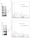
Mass spectrometry of peptide pair derived from CYP17A1 cross-linked with b5. A, EDC cross-linking of wild-type CYP17A1 and b5 followed by SDS-PAGE analysis with Denville blue staining. Reactions were performed as described in Fig. 3, and the entire reaction mixture was loaded on the gel. B, representative high resolution fragmentation spectrum of cross-linked peptide between CYP17A1 and b5 identified by StavroX: CYP17A1-WT (84EVLIKK89) and b5 (53EQAGGDATENFEDVGHSTDAR73). The numbers in parentheses indicate the positions of the cross-linked peptide sequences with the cross-linked residues underlined. Signals of y-type ions are shown in blue, and signals of b-type ions are shown in red. The measurement error of the ion is 0.3 ppm. C, EDC cross-linking of R358K and b5 followed by SDS-PAGE and Denville blue staining. D, representative high resolution fragmentation spectrum of cross-linked peptide pair between CYP17A1-R358K and b5: CYP17A1-R358K (84EVLIKK89) and b5 (53EQAGGDATENFEDVGHSTDAR73). Residue numbering and spectrum annotation are the same as in A and B. The measurement error of the ion is 1.5 ppm.
FIGURE 5.
FIGURE 5.
Mass spectrometry of peptide pair derived from CYP17A1 mutation R347K cross-linked with b5. A, EDC cross-linking of CYP17A1 mutation R347K and b5 followed by SDS-PAGE analysis with Denville blue staining. Reactions were performed as described in Fig. 3, and the entire reaction mixture was loaded on the gel. B, representative high resolution fragmentation spectrum of cross-linked peptide between CYP17A1-R347K and b5 identified by StavroX: R347K (341TPTISDKNR349) and b5 (40FLEEHPGGEEVLR52). The numbers in parentheses indicate the positions of the cross-linked peptide sequences with the cross-linked residues underlined. Signals of y-type ions are shown in blue, and signals of b-type ions are shown in red. The measurement error of the ion is 1.6 ppm.
FIGURE 6.
FIGURE 6.
Docking model of CYP17A1 interaction with b5, based on crystal structures of CYP17A1 (PDB code 3RUK) and b5 (PDB code 2I96). The docking was performed using the programs HADDOCK with the distance constraints of two intermolecular cross-links, Lys-88(CYP17A1)-Glu-61(b5) and Arg-347(CYP17A1)-Glu-42(b5), as well as Glu-48 and Glu-49 of b5. A, interaction of amino acid Lys-88 (red) in CYP17A1 and Glu-61 (blue) of b5 are shown on a surface model. Glu-49 of b5 in the center of complex is shown in blue. B, schematic model of CYP17A1 interacting with b5. The heme groups in CYP17A1 and b5 are shown in red and orange sticks, respectively, and the abiraterone molecule in the crystal structure is shown in green. The central I-helix is displayed in purple, and amino acids Lys-88, Arg-347, and Arg-358 of CYP17A1, which interact with b5, are shown in magenta. The side chain of glutamic acids 42, 48, and 49 of b5 are shown in blue. C, close-up view of the CYP17A1-b5 interaction interface is shown, where the glutamic acid residues in b5 interacting with CYP17A1 are visible. D, CYP17A1-b5 interactions were detected using the PISA server and presented as a space-filled model. Interface residues at less than 3.9 Å from interaction sites are shown in red (including Arg-347, Arg-358, and Arg-449) for CYP17A1 and green (including Glu-42, Glu-48, Glu-49, and Arg-52) for b5, respectively. The buried residues were colored in gray, and the rest of the residues are shown in blue for CYP17A1 and cyan for b5.

References

    1. Miller W. L., Auchus R. J. (2011) The molecular biology, biochemistry, and physiology of human steroidogenesis and its disorders. Endocr. Rev. 32, 81–151 - PMC - PubMed
    1. Attard G., Reid A. H., Yap T. A., Raynaud F., Dowsett M., Settatree S., Barrett M., Parker C., Martins V., Folkerd E., Clark J., Cooper C. S., Kaye S. B., Dearnaley D., Lee G., de Bono J. S. (2008) Phase I clinical trial of a selective inhibitor of CYP17, abiraterone acetate, confirms that castration-resistant prostate cancer commonly remains hormone driven. J. Clin. Oncol. 26, 4563–4571 - PubMed
    1. Mostaghel E. A., Nelson P. S. (2008) Intracrine androgen metabolism in prostate cancer progression: mechanisms of castration resistance and therapeutic implications. Best Pract. Res. Clin. Endocrinol. Metab. 22, 243–258 - PMC - PubMed
    1. Miller W. L., Auchus R. J., Geller D. H. (1997) The regulation of 17,20 lyase activity. Steroids 62, 133–142 - PubMed
    1. Auchus R. J., Lee T. C., Miller W. L. (1998) Cytochrome b5 augments the 17,20-lyase activity of human P450c17 without direct electron transfer. J. Biol. Chem. 273, 3158–3165 - PubMed

Publication types

MeSH terms

LinkOut - more resources