

Comparative Study
doi: 10.1186/s12974-014-0170-y.
Immune responses in rapidly progressive dementia: a comparative study of neuroinflammatory markers in Creutzfeldt-Jakob disease, Alzheimer's disease and multiple sclerosis
- PMID: 25315814
- PMCID: PMC4207356
- DOI: 10.1186/s12974-014-0170-y
Item in Clipboard
Comparative Study
Immune responses in rapidly progressive dementia: a comparative study of neuroinflammatory markers in Creutzfeldt-Jakob disease, Alzheimer's disease and multiple sclerosis
J Neuroinflammation.
.
Abstract
Immunological responses may contribute to disease progression and clinical heterogeneity in neurodegenerative dementia, for example, Alzheimer's disease (AD) and Creutzfeldt-Jakob disease (CJD). Recently, a rapidly progressive form of AD (rpAD) has been described. On neuropathological grounds classical AD and rpAD are not distinguishable at present. All those protein aggregopathies show a state of chronic inflammation with microglia activation and production of proinflammatory cytokines. In this context, it is hypothesized that the severity of the surrounding inflammation substantially contributes to disease progression and accelerated disease courses as seen in rpAD.
Figures
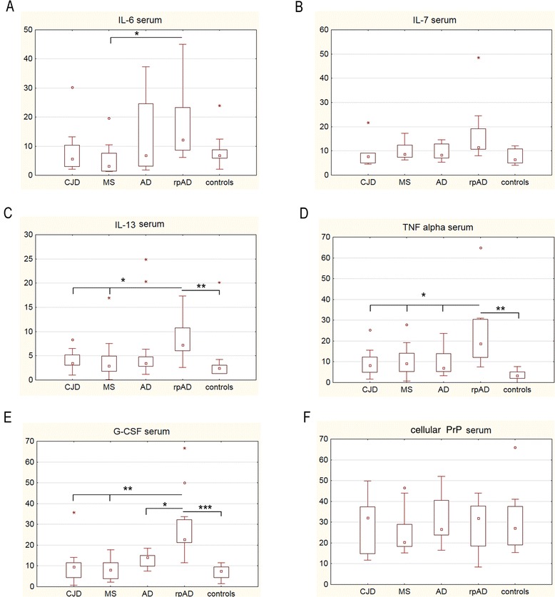
Different immune responses in the serum of patients with rapidly progressive dementia.
(A-E) Profiling of cytokines (measured in pg/ml) in serum from Creutzfeldt-Jakob disease (CJD) (n =12), multiple sclerosis (MS) (n =12), Alzheimer’s disease (AD) (n =20), a rapidly progressive form of AD (rpAD) (n =15) and control (n =12) patients was performed using the Bio-Plex Pro human cytokine 17-plex assay. A significant distinction in rpAD patients was identified for IL-6, IL-13, TNF-α and G-CSF. (F) PrPC level in serum did not vary among groups. Columns represent means with SD. Statistics were performed by using the non-parametric Kruskal-Wallis test (+Tukey’s post hoc tests). The number of stars indicates the significance level: one star (*) for P <0.05, two (**) for P <0.01 and three (***) for P <0.001.
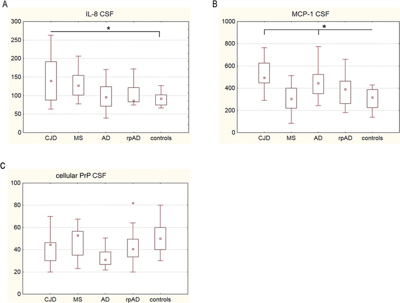
Different immune responses in cerebrospinal fluid (CSF) of patients with rapidly progressive dementia.
(A-B) Profiling of cytokines (pg/ml) in CSF from Creutzfeldt-Jakob disease (CJD) (n =12), multiple sclerosis (MS) (n =12), Alzheimer’s disease (AD) (n =20), a rapidly progressive form of AD (rpAD) (n =15) and control (n =12) patients was performed by using the Bio-Plex Pro human cytokine 17-plex assay. A significant increase of IL-8 was found in CJD patients, while the level of MCP-1 was significantly elevated in CJD and AD patients but not in rpAD patients compared to control donors. (C) PrPC level in the CSF did not vary significantly among groups (n =12). Columns represent means with SD. Statistics were performed by using the non-parametric Kruskal-Wallis test (+ Turkey's post hoc tests). The number of stars indicates the significance level: one star (*) for P <0.05, two (**) for P <0.01 and three (***) for P <0.001.
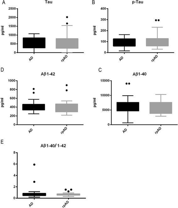
No differences in the levels of cerebrospinal fluid (CSF) dementia markers between patients with Alzheimer’s disease (AD) and patients with a rapidly progressive form of Alzheimer’s disease (rpAD).
(A-D) Expression levels of total Tau, p-Tau, Aβ1-40 and Aβ1-42 in the CSF of AD and rpAD (each, n =8) were measured by ELISA. (E) Aβ ratios were calculated from Aβ1-40 and Aβ1-42. No significant variations could be detected in either cohort. Error bars represent means with SD. The number of stars indicates the significance level: one star (*) for P <0.05, two (**) for P <0.01 and three (***) for P <0.001.
References
-
- Akiyama H, Barger S, Barnum S, Bradt B, Bauer J, Cole GM, Cooper NR, Eikelenboom P, Emmerling M, Fiebich BL, Finch CE, Frautschy S, Griffin WS, Hampel H, Hull M, Landreth G, Lue L, Mrak R, Mackenzie IR, McGeer PL, O’Banion MK, Pachter J, Pasinetti G, Plata-Salaman C, Rogers J, Rydel R, Shen Y, Streit W, Strohmeyer R, Tooyoma I, et al. Inflammation and Alzheimer’s disease. Neurobiol Aging. 2000;21:383–421. doi: 10.1016/S0197-4580(00)00124-X. - DOI - PMC - PubMed
-
- Hoozemans JJM, Rozemuller AJM, Janssen I, De Groot CJA, Veerhuis R, Eikelenboom P. Cyclooxygenase expression in microglia and neurons in Alzheimer’s disease and control brains. Acta Neuropathol (Berl) 2001;101:2–8. - PubMed
Publication types
MeSH terms
Substances
LinkOut - more resources
Full Text Sources
Other Literature Sources
Medical
