. 2014 Dec 12;426(24):3946-3959.
doi: 10.1016/j.jmb.2014.10.008. Epub 2014 Oct 12.

GEN1 from a thermophilic fungus is functionally closely similar to non-eukaryotic junction-resolving enzymes

Affiliations

GEN1 from a thermophilic fungus is functionally closely similar to non-eukaryotic junction-resolving enzymes

Alasdair D J Freeman et al. J Mol Biol. .

Abstract

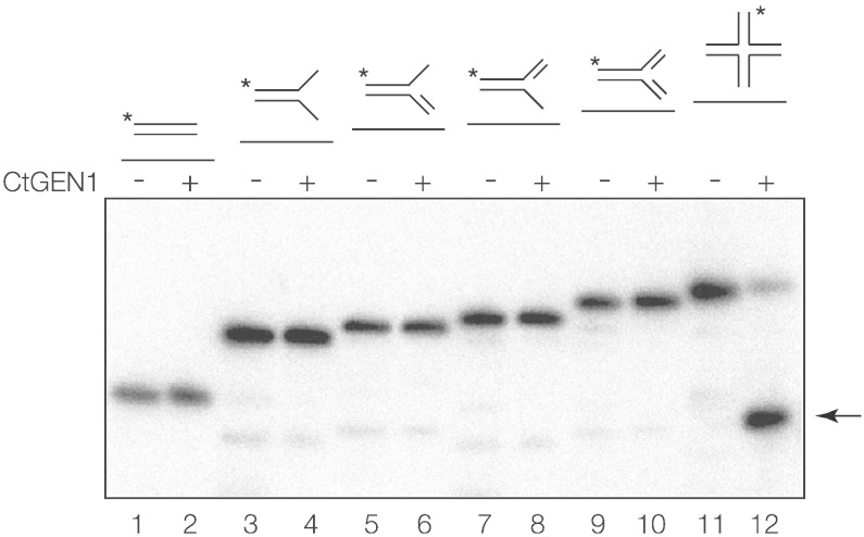
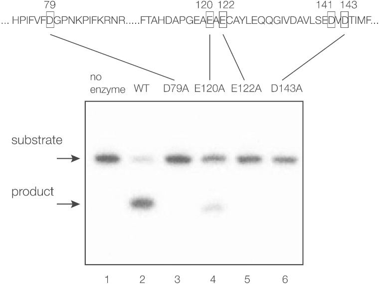
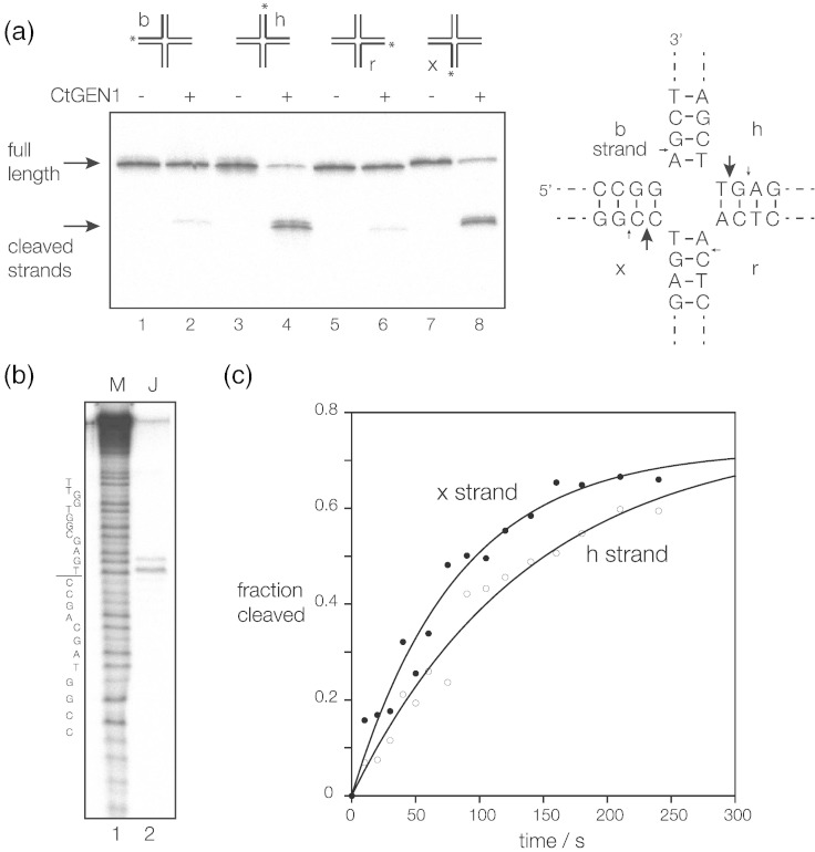
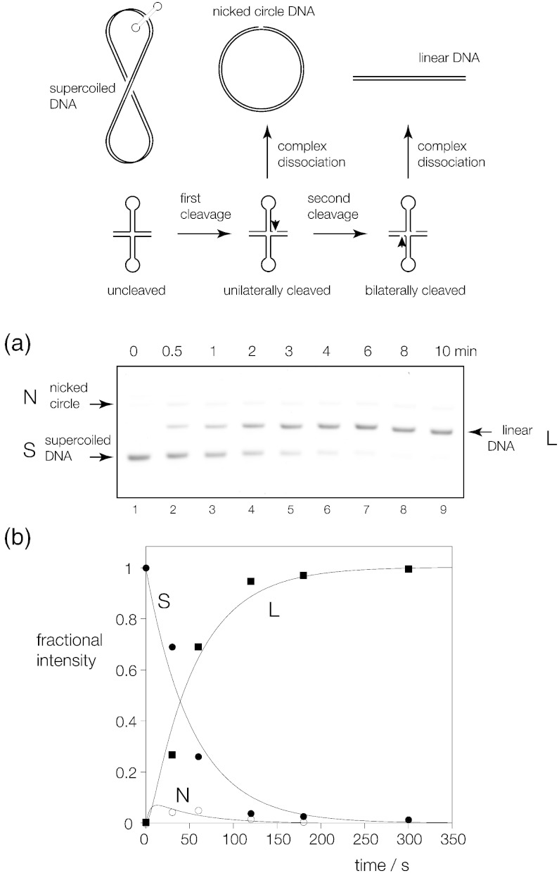
Processing of Holliday junctions is essential in recombination. We have identified the gene for the junction-resolving enzyme GEN1 from the thermophilic fungus Chaetomium thermophilum and expressed the N-terminal 487-amino-acid section. The protein is a nuclease that is highly selective for four-way DNA junctions, cleaving 1nt 3' to the point of strand exchange on two strands symmetrically disposed about a diagonal axis. CtGEN1 binds to DNA junctions as a discrete homodimer with nanomolar affinity. Analysis of the kinetics of cruciform cleavage shows that cleavage of the second strand occurs an order of magnitude faster than the first cleavage so as to generate a productive resolution event. All these properties are closely similar to those described for bacterial, phage and mitochondrial junction-resolving enzymes. CtGEN1 is also similar in properties to the human enzyme but lacks the problems with aggregation that currently prevent detailed analysis of the latter protein. CtGEN1 is thus an excellent enzyme with which to engage in biophysical and structural analysis of eukaryotic GEN1.

Keywords: Chaetomium thermophilum; DNA recombination and repair; FEN1; Holliday junction resolution; thermophilic proteins.


Figures

None
Graphical abstract
Fig. 1
Fig. 1
Identification and expression of a putative GEN1 sequence from C. thermophilum. (a ) Alignment of the protein sequences of human (Hs) FEN1 and GEN1 and the putative C. thermophilum (Ct) GEN1, shaded by identity. Seven conserved acidic amino acids involved in metal ion binding in the active site of FEN1 are boxed red, and the conserved lysine is boxed blue. (b) Expression of CtGEN11–487. The protein fragment was expressed in E. coli as a fusion with GFP that was subsequently cleaved using TEV protease. Purified protein was analyzed by electrophoresis in 10% polyacrylamide containing SDS (Fisher). Tracks: 1, a mixture of proteins as a size marker, with molecular mass (kDa) written on the left; 2, the CtGEN11–487-GFP fusion protein; 3, purified CtGEN11–487; 4, GFP released from the fusion.
Fig. 2
Fig. 2
Substrate selectivity for CtGEN11–487. A number of branched DNA species were prepared, with one strand radioactively 5′-32P-labeled (indicated by an asterisk). Each was incubated with (even tracks) or without (odd tracks) 50 nM CtGEN11–487 in 10 mM Hepes (pH 7.5) in the presence of 10 mM MgCl2. Potential products were separated by electrophoresis in 6% polyacrylamide and visualized by phosphorimaging. The substrates were duplex (tracks 1 and 2), splayed duplex (tracks 3 and 4), 3′ flap (tracks 5 and 6), 5′ flap (tracks 7 and 8), nicked three-way junction (tracks 9 and 10) and four-way junction Jbm5 (tracks 11 and 12). Only the four-way junction was significantly cleaved, with the product arrowed.
Fig. 3
Fig. 3
Cleavage activity on a DNA junction by putative active-site mutants of CtGEN11–487. Conserved acidic residues were individually mutated to alanine and the cleavage activity against junction 3 was tested using purified CtGEN11–487-GFP fusions. Junction 3 radioactively 5′-32P-labeled on the x strand was incubated without enzyme (track 1) or with wild type (track 2), D79A (track 3), E120A (track 4), E122A (track 5) or D143A (track 6) GEN1 for 4 min at 37 °C, and any products of cleavage were separated by electrophoresis in a 15% polyacrylamide and visualized by phosphorimaging.
Fig. 4
Fig. 4
Location and rates of cleavage of the four strands of a four-way junction. Four versions of a junction with arms of 25 bp and a core sequence corresponding to junction 3 were each radioactively 5′-32P-labeled on a single strand. Each was incubated with 100 nM CtGEN11–487 in 10 mM Hepes (pH 7.5), 10 mM MgCl2, 50 mM NaCl, 0.1% BSA and 1 mM DTT at 37 °C for 10 min, and the products were separated by electrophoresis in a 15% polyacrylamide and visualized by phosphorimaging. (a) Phosphorimage of the gel. Samples were applied in the order b, h, r and x strands labeled, with even- and odd-numbered tracks containing samples with and without enzyme added. All strands were cleaved to some degree, but the h and x strands were cleaved more strongly than the b or r strands. (b) The product of h-strand cleavage by CtGEN11–487 was electrophoresed in a 15% polyacrylamide sequencing gel in TBE containing 8 M urea alongside a sequence ladder derived from partial chemical degradation of the h strand in order to map the positions of cleavage at nucleotide resolution. The sequence of the h strand is shown on the left, with the position of strand exchange indicated by the line. The positions of cleavage in all four strands are shown on the schematic of the central sequence of the junction, with the larger arrows indicating strong cleavage. (c) Reaction progress plotted as a function of time for the h (open circles) and x (filled circles) strands. The data have been fitted to single exponential functions (lines).
Fig. 5
Fig. 5
Binding of CtGEN11–487 to junction 3. Junction 3 was prepared with 25 bp arms, radioactively 5′-32P-labeled on the x strand. We incubated 82 pM junction with increasing concentrations of CtGEN11–487 in 10 mM Hepes (pH 7.5), 50 mM NaCl, 0.1% BSA and 1 mM DTT with either 1 mM EDTA or 1 mM Ca2 + for 60 min and electrophoresed in 6% polyacrylamide under non-denaturing conditions. (a and b) Phosphorimages of the gels electrophoresed in (a) 1 mM EDTA and (b) 1 mM Ca2 +. Tracks contain DNA junction incubated with increasing concentrations of CtGEN11–487 left to right. The concentration of CtGEN11–487 is shown over each track. DNA–protein complexes are visible migrating more slowly than the free junction (arrowed right). A complex of intermediate mobility is visible in EDTA, but in Ca2 +, essentially only a single complex is evident. (c) The fractional intensity of the retarded complex in Ca2 + is plotted as a function of CtGEN11–487 concentration (filled circles). The data have been fitted to two models. A standard binding isotherm (line) gives an affinity Kd = 10 nM, but the data clearly exhibit cooperativity. They have therefore additionally been fitted to the Hill equation (broken line).
Fig. 6
Fig. 6
CtGEN11–487 binds to a DNA junction as dimer. Complexes have been formed with CtGEN11–487 alone and fused to GFP. These have been analyzed by gel electrophoresis in 6% polyacrylamide. In each case, free junction was electrophoresed in track 1. The experiment was performed in two ways. (a) We incubated 160 nM junction with 40 nM CtGEN11–487 in 10 mM Hepes (pH 7.5), 50 mM NaCl, 0.1% BSA, 1 mM DTT and 1 mM CaCl2 (track 2), with increasing concentrations of CtGEN1-GFP fusion (tracks 3–7; concentrations indicated above each track). (b) We incubated 80 nM junction with 40 nM CtGEN1-GFP fusion in 10 mM Hepes (pH 7.5), 50 mM NaCl, 0.1% BSA, 1 mM DTT and 1 mM CaCl2 (track 2), with increasing concentrations of CtGEN11–487 fusion (tracks 3–7; concentrations indicated above each track). Note that, in each case, three retarded complexes are observed, consistent with CtGEN11–487 binding as a dimer to the DNA junction.
Fig. 7
Fig. 7
Analysis of bilateral cleavage in a DNA junction using a supercoil-stabilized cruciform substrate. The principle of the experiment is shown in the scheme (top). A cruciform structure contains a four-way junction that is a substrate for resolving enzymes. However, the cruciform requires negative supercoiling for stabilization. Unilateral cleavage of the junction followed by dissociation of the protein leads to the formation of a nicked circle in which the cruciform substrate is no longer present. By contrast, subsequent second cleavage within the lifetime of the complex generates a linear product as the cruciform is now bilaterally cleaved. (a) The supercoiled DNA substrate and the nicked and linear products are readily separated by electrophoresis in 1% agarose. Supercoiled plasmid pBHR3 was incubated with 200 nM CtGEN11–487 in 10 mM Hepes (pH 7.5), 50 mM NaCl, 1 mM DTT and 0.1% BSA at 37 °C. After the cleavage reaction was initiated by addition of MgCl2 to 10 mM, samples were removed at different times, protein was removed by treatment with proteinase K and electrophoresed in a 1% agarose gel and DNA was fluorescently stained. A fluoroimage is shown. With time, the supercoiled DNA is converted to linear product, with a low intensity of nicked circular DNA appearing as a transient intermediate. (b) The intensities of the three species were quantified and plotted as a function of time (lower). These data are fitted to the integrated rate Eqs. (3), (4), (5) shown in the main text.

Similar articles

Cited by

References

    1. Holliday R. A mechanism for gene conversion in fungi. Genet Res. 1964;5:282–304. - PubMed
    1. Ellis N.A., Groden J., Ye T.Z., Straughen J., Lennon D.J., Ciocci S. The Bloom's syndrome gene product is homologous to RecQ helicases. Cell. 1995;83:655–666. - PubMed
    1. Wu L., Hickson I.D. The Bloom's syndrome helicase suppresses crossing over during homologous recombination. Nature. 2003;426:870–874. - PubMed
    1. Cejka P., Plank J.L., Bachrati C.Z., Hickson I.D., Kowalczykowski S.C. Rmi1 stimulates decatenation of double Holliday junctions during dissolution by Sgs1-Top3. Nat Struct Mol Biol. 2010;17:1377–1382. - PMC - PubMed
    1. Bloom D. Congenital telangiectatic erythema resembling lupus erythematosus in dwarfs; probably a syndrome entity. AMA Am J Dis Child. 1954;88:754–758. - PubMed

Publication types

MeSH terms