. 2014 Oct;198(2):723-33.
doi: 10.1534/genetics.114.168211.

A novel ribosomopathy caused by dysfunction of RPL10 disrupts neurodevelopment and causes X-linked microcephaly in humans

Affiliations

A novel ribosomopathy caused by dysfunction of RPL10 disrupts neurodevelopment and causes X-linked microcephaly in humans

Susan S Brooks et al. Genetics. 2014 Oct.

Abstract

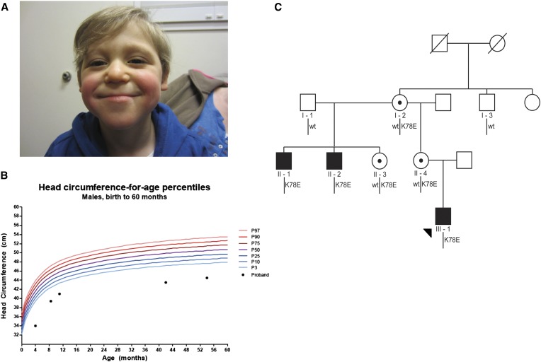
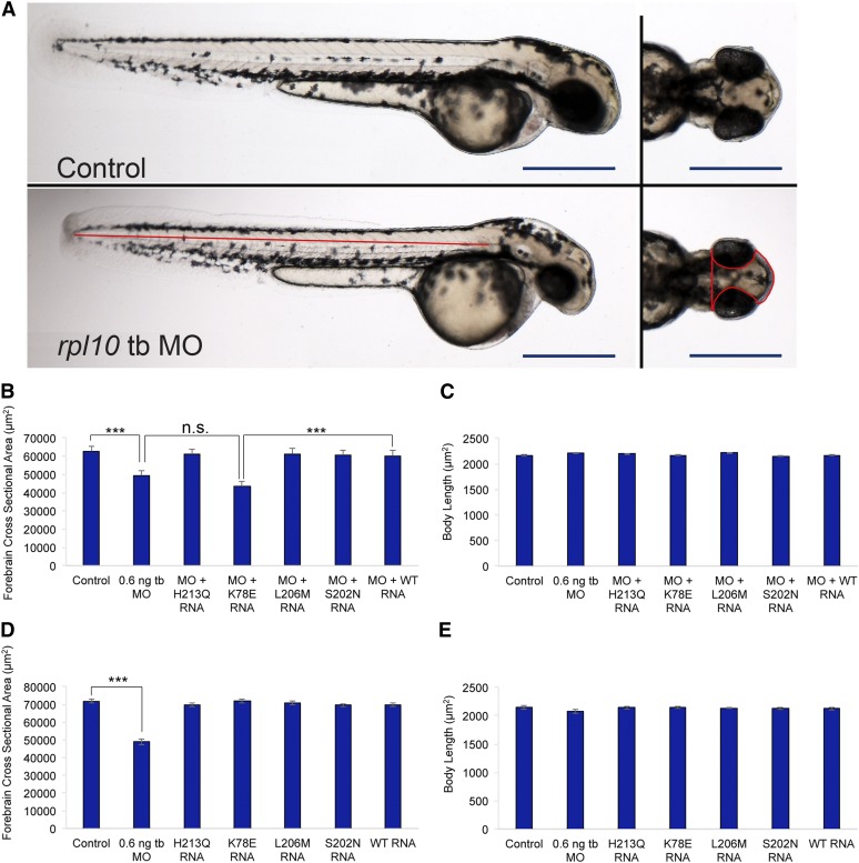
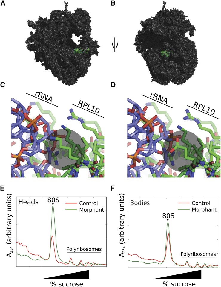
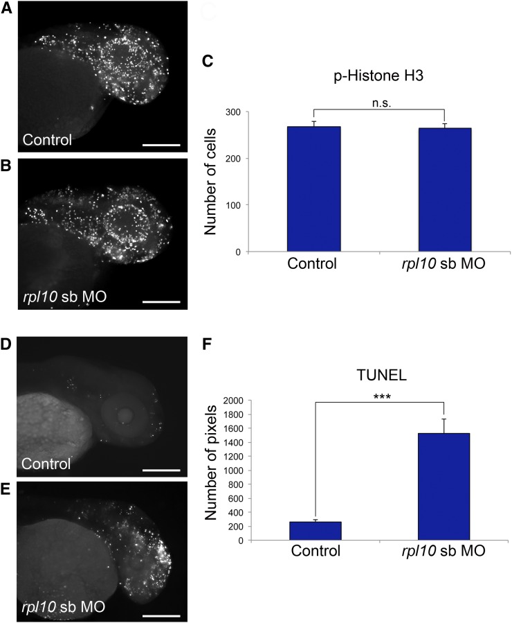
Neurodevelopmental defects in humans represent a clinically heterogeneous group of disorders. Here, we report the genetic and functional dissection of a multigenerational pedigree with an X-linked syndromic disorder hallmarked by microcephaly, growth retardation, and seizures. Using an X-linked intellectual disability (XLID) next-generation sequencing diagnostic panel, we identified a novel missense mutation in the gene encoding 60S ribosomal protein L10 (RPL10), a locus associated previously with autism spectrum disorders (ASD); the p.K78E change segregated with disease under an X-linked recessive paradigm while, consistent with causality, carrier females exhibited skewed X inactivation. To examine the functional consequences of the p.K78E change, we modeled RPL10 dysfunction in zebrafish. We show that endogenous rpl10 expression is augmented in anterior structures, and that suppression decreases head size in developing morphant embryos, concomitant with reduced bulk translation and increased apoptosis in the brain. Subsequently, using in vivo complementation, we demonstrate that p.K78E is a loss-of-function variant. Together, our findings suggest that a mutation within the conserved N-terminal end of RPL10, a protein in close proximity to the peptidyl transferase active site of the 60S ribosomal subunit, causes severe defects in brain formation and function.

Keywords: X-linked; microcephaly; ribosome; translational elongation; zebrafish.


Figures

Figure 1
Figure 1
Microcephaly in an X-linked pedigree harboring RPL10 p.K78E. (A) The proband (III-1) at age 41 months. He displays microcephaly, a thin upper lip, and mandibular prognathism (see Table 1 for full phenotypic description). (B) Head circumference chart for the proband. Microcephaly was of prenatal onset and growth continued to follow a normal curve at this reduced trajectory. (C) X-linked segregation of RPL10 c.232A > G; p.K78E in a three-generation pedigree. Subsequent to identification of p.K78E in individual II-1 by a next-generation sequencing X-linked intellectual disability diagnostic panel, Sanger sequencing confirmed segregation of the mutation in three affected males and three carrier females. A healthy maternal great uncle (I-3) of the proband was wild type at this locus, and both carrier mothers (I-2 and II-4) displayed fully skewed X inactivation.
Figure 2
Figure 2
Suppression of rpl10 in zebrafish results in reduced head size and p.K78E is a loss-of-function variant. (A) Live larval images of control (top) and rpl10 tb-MO-injected embryos (bottom). Left panels show lateral views of whole larvae with similar body lengths; right panels show dorsal views showing a reduced head size in morphants. Red lines indicate head size measurements quantified in B and D and body length measurements quantified in C and E. Bars, 500 μm. (B) Quantification of head area for rpl10 MO and MO co-injected with human RPL10 mRNA. (C) Quantification of body length for rpl10 MO and MO co-injected with human RPL10 mRNA. (D) Quantification of head size for embryos injected with RPL10 mRNA alone. (E) Quantification of body length for embryos injected with RPL10 mRNA alone. Head area and body length measurements were carried out at 2 days postfertilization (dpf), using embryos injected with 0.6 ng tb-MO and/or 50 pg mRNA; n = 30 for each injection batch with masked scoring were repeated with similar results. Error bars indicate standard error of the mean (SEM). ***P < 0.0001 (two-tailed t-test comparisons between MO-injected and rescued embryos).
Figure 3
Figure 3
rpl10 morphants display reduced bulk translation, especially in larval heads, as indicated by polyribosome structure. (A) Rendering of RPL10 in green on a eukaryotic ribosome in proximity to the peptidyl transferase active site. Protein Data Bank (PDB) entries 4a17, 4a19, and 2xzm were merged using a yeast ribosome (PDBs 2xzm and 3o58) as a guide. (B) As in A, rotated 90°. (C) Interaction of wild-type RPL10 K78 with the ribosomal (r)RNA. K78 interacts mostly with the negatively charged 28S rRNA and is indicated by a gray circle. (D) Simulation of the K78E mutation, indicated by a gray circle. (E) Sucrose gradient analysis of polyribosome structure in heads of 5 day postfertilization (dpf) rpl10 morphants and controls. Note the increase in 80S abundance with a corresponding decrease in polyribosomes for morphants vs. controls, indicative of a decrease in translational activity. (F) Sucrose gradient analysis of bodies of rpl10 morphants and controls. Polyribosome profiles are similar.
Figure 4
Figure 4
rpl10 morphants display normal cell proliferation and increased apoptosis in the brain. (A and B) Whole-mount phospho-histone H3 staining for proliferating cells (M-phase marker) in control and rpl10 morphants at 2 dpf (lateral views). (C) Quantification of phospho-histone H3-positive cells from 20 embryos each (control embryos or embryos injected with rpl10 MO). Data are represented as the mean ± SEM. n.s., nonsignificant (two-tailed t-test comparisons between sb-MO-injected and rescued embryos). (D and E) TUNEL staining for apoptotic cells in control and rpl10 morphants at 2 dpf (lateral views). (F) Quantification of TUNEL staining intensities from 20 embryos each (control embryos or embryos injected with rpl10 sb-MO). Data are represented as the mean ± SEM. ***P < 0.0001 (two-tailed t-test comparisons between MO-injected and rescued embryos); similar results were obtained with the rpl10 tb-MO. Bars, 250 μm.

Comment in

References

    1. Adzhubei I. A., Schmidt S., Peshkin L., Ramensky V. E., Gerasimova A., et al. , 2010. A method and server for predicting damaging missense mutations. Nat. Methods 7: 248–249 - PMC - PubMed
    1. Aspesi A., Pavesi E., Robotti E., Crescitelli R., Boria I., et al. , 2014. Dissecting the transcriptional phenotype of ribosomal protein deficiency: implications for Diamond-Blackfan anemia. Gene 545: 282–289 - PMC - PubMed
    1. Ben-Shem A., Garreau de Loubresse N., Melnikov S., Jenner L., Yusupova G., et al. , 2011. The structure of the eukaryotic ribosome at 3.0 A resolution. Science 334: 1524–1529 - PubMed
    1. Beunders G., Voorhoeve E., Golzio C., Pardo L. M., Rosenfeld J. A., et al. , 2013. Exonic deletions in AUTS2 cause a syndromic form of intellectual disability and suggest a critical role for the C terminus. Am. J. Hum. Genet. 92: 210–220 - PMC - PubMed
    1. Bicknell L. S., Walker S., Klingseisen A., Stiff T., Leitch A., et al. , 2011. Mutations in ORC1, encoding the largest subunit of the origin recognition complex, cause microcephalic primordial dwarfism resembling Meier-Gorlin syndrome. Nat. Genet. 43: 350–355 - PubMed

Publication types