. 2014 Oct 28;111(43):15438-43.
doi: 10.1073/pnas.1412767111. Epub 2014 Oct 14.

Direct activation of RIP3/MLKL-dependent necrosis by herpes simplex virus 1 (HSV-1) protein ICP6 triggers host antiviral defense

Affiliations

Direct activation of RIP3/MLKL-dependent necrosis by herpes simplex virus 1 (HSV-1) protein ICP6 triggers host antiviral defense

Xing Wang et al. Proc Natl Acad Sci U S A. .

Abstract

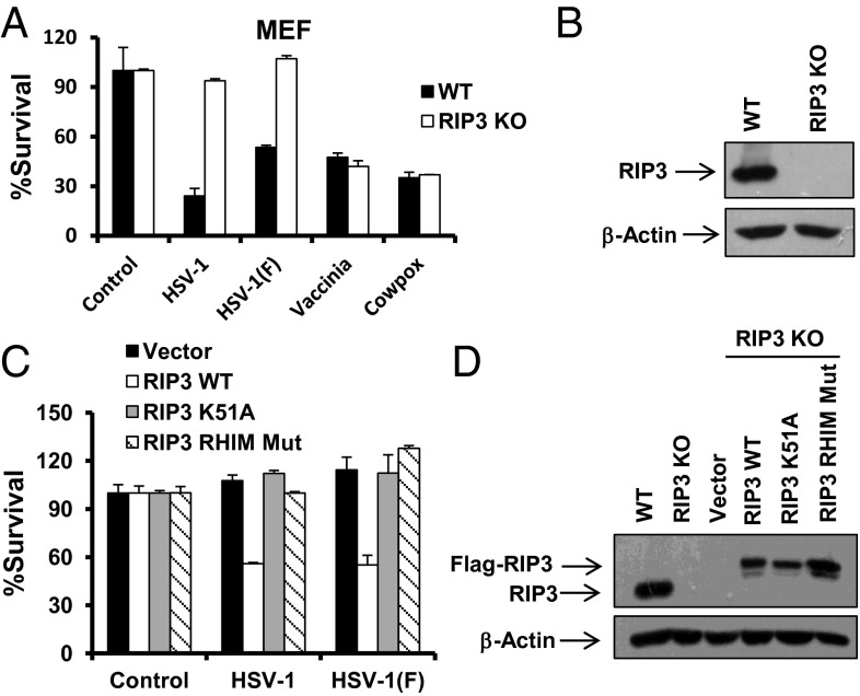
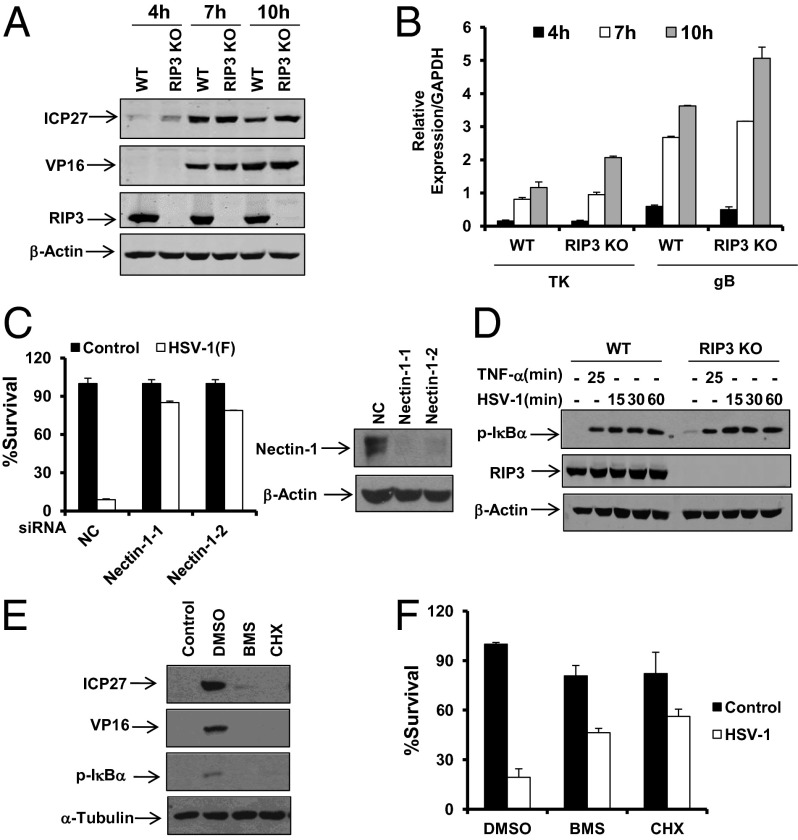
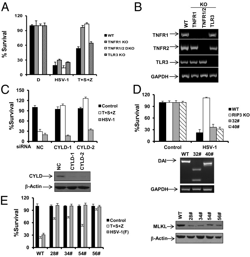
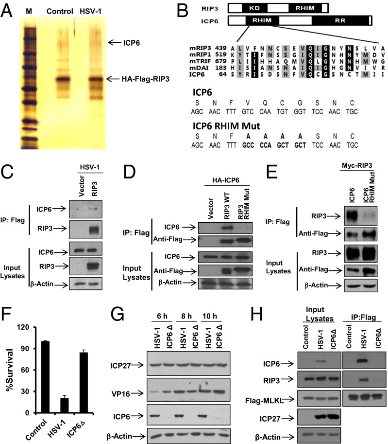
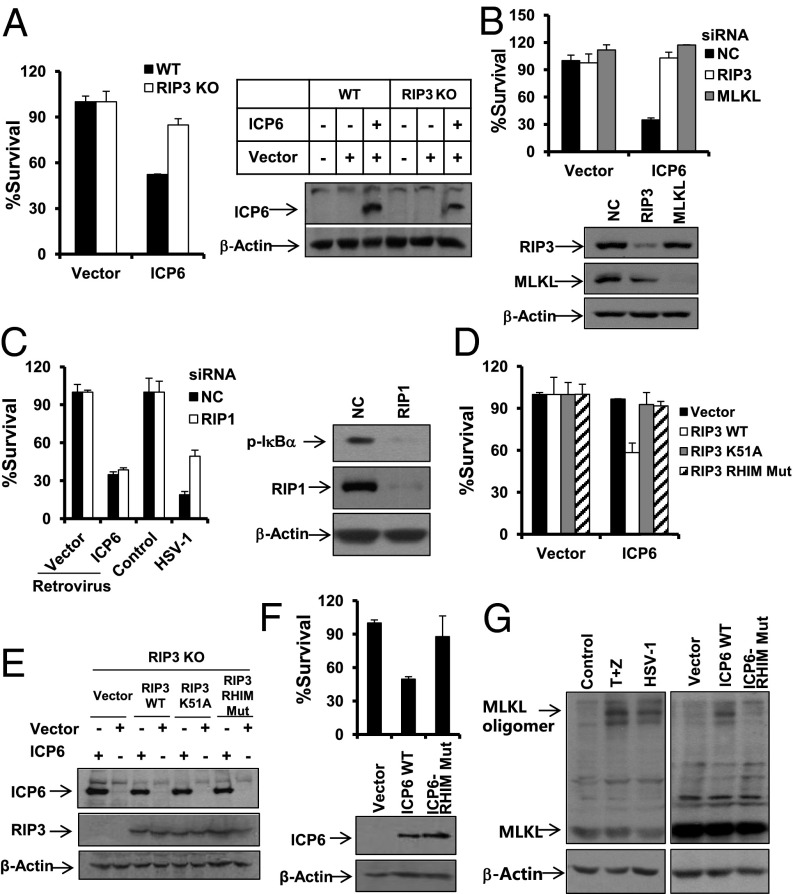
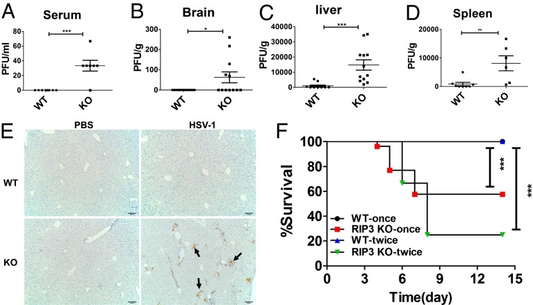
The receptor-interacting kinase-3 (RIP3) and its downstream substrate mixed lineage kinase domain-like protein (MLKL) have emerged as the key cellular components in programmed necrotic cell death. Receptors for the cytokines of tumor necrosis factor (TNF) family and Toll-like receptors (TLR) 3 and 4 are able to activate RIP3 through receptor-interacting kinase-1 and Toll/IL-1 receptor domain-containing adapter inducing IFN-β, respectively. This form of cell death has been implicated in the host-defense system. However, the molecular mechanisms that drive the activation of RIP3 by a variety of pathogens, other than the above-mentioned receptors, are largely unknown. Here, we report that human herpes simplex virus 1 (HSV-1) infection triggers RIP3-dependent necrosis. This process requires MLKL but is independent of TNF receptor, TLR3, cylindromatosis, and host RIP homotypic interaction motif-containing protein DNA-dependent activator of IFN regulatory factor. After HSV-1 infection, the viral ribonucleotide reductase large subunit (ICP6) interacts with RIP3. The formation of the ICP6-RIP3 complex requires the RHIM domains of both proteins. An HSV-1 ICP6 deletion mutant failed to cause effective necrosis of HSV-1-infected cells. Furthermore, ectopic expression of ICP6, but not RHIM mutant ICP6, directly activated RIP3/MLKL-mediated necrosis. Mice lacking RIP3 exhibited severely impaired control of HSV-1 replication and pathogenesis. Therefore, this study reveals a previously uncharacterized host antipathogen mechanism.

Keywords: HSV-1; ICP6; MLKL; RIP3; programmed necrosis.


Conflict of interest statement

The authors declare no conflict of interest.

Figures

Fig. 1.
Fig. 1.
HSV-1 infection activates RIP3-dependent necrosis. (A) WT and RIP3 KO MEFs were infected with the HSV-1 or HSV-1 F strain at a multiplicity of infection (MOI) of 5 for around 15 h, or infected with vaccinia or cowpox virus for around 28 h. Identical MOI was used in MEF in later experiments unless otherwise stated. The cell-survival rate was determined by measuring ATP levels. (B) Cell lysates collected from WT or RIP3 KO MEFs were subjected to Western-blot analysis. (C) RIP3 KO MEFs expressing WT RIP3 (RIP3 WT) or RIP3 kinase dead mutant (RIP3 K51A) or RHIM domain mutant form of RIP3 (RIP3 RHIM Mut) were infected with HSV-1 or F strain for 16–18 h. The cell-survival rate was determined by measuring ATP levels. (D) Cell lysates collected from RIP3 KO MEFs expressing RIP3 WT or RIP3 K51A or RIP3 RHIM Mut were subjected to Western-blot analysis.
Fig. 2.
Fig. 2.
RIP3 is dispensable for HSV-1 entry, replication, and virus-induced NF-κB activation. (A and B) WT and RIP3 KO MEFs were infected with HSV-1 (MOI = 2) for the indicated time. Cell lysates were then collected and subjected to Western-blot analysis for the indicated proteins (A). Quantitative PCR (qPCR) was performed to measure the expression levels of thymidine kinase (TK) and gB (B). The error bars show the SD from duplicate qPCR reactions. (C) MEFs were transfected with negative control (NC) or nectin-1 siRNA oligos for 48 h. Cells were treated as indicated for an additional 16–18 h. The cell-survival rate was determined by measuring ATP levels. Cell lysates were collected 48 h posttransfection and subjected to Western-blot analysis. (D) WT and RIP3 KO MEFs were treated with TNF-α for 25 min or infected with HSV-1 (MOI = 2). At the indicated time points, cell lysates were collected and subjected to Western-blot analysis. (E) MEFs were treated with DMSO, BMS-345541 (BMS), or Cycloheximide (CHX) for 1 h before infection with HSV-1 (MOI = 2) for an additional 8 h. Cell lysates were then collected and subjected to Western-blot analysis. BMS, 20 µM; CHX, 5 µg/mL. (F) MEFs were treated with DMSO, BMS, or CHX for 1 h before infection with HSV-1 for an additional 16 h. The cell-survival rate was determined by measuring ATP levels.
Fig. 3.
Fig. 3.
HSV-1 infection-induced necrosis requires MLKL but is independent of TNFR, TLR3, CYLD, and DAI. (A) MEFs isolated from WT, TNFR1 KO, TNFR1/TNFR2 double KO, and TLR3 KO mice were treated as indicated for 16–18 h. The cell survival rate was determined by measuring ATP levels. T, TNF-α; S, Smac mimetic; Z, z-VAD. (B) Total mRNA was collected from the indicated MEFs. The mRNA expression levels of the indicated genes were measured. (C) MEFs were transfected with NC or CYLD siRNA oligos for 48 h. Cells were treated as indicated for an additional 16–18 h. The cell-survival rate was determined by measuring ATP levels. Cell lysates were collected 48 h posttransfection and subjected to Western-blot analysis. (D) WT MEFs, RIP3 KO MEFs, or two DAI KO clones (32# and 40#) were infected with HSV-1 for 16–18 h. The cell-survival rate was determined by measuring ATP levels. Genomic DNA was collected from DAI KO clone 32# or 40#, and alterations of DAI DNA sequence were analyzed by PCR. (E) MEFs or MLKL-shRNA stable cell lines as indicated were infected with the HSV-1 F strain for 16–18 h. Cell lysates were collected and subjected to Western-blot analysis. The cell-survival rate was determined by measuring ATP levels. MLKL-shRNA, MEFs stably expressing a shRNA targeting mouse MLKL.
Fig. 4.
Fig. 4.
Virus protein ICP6 interacts with RIP3 through the RHIM domains of both proteins and is required for HSV-1–induced necrosis. (A) RIP3 KO MEFs expressing WT RIP3 (RIP3 was double tagged with Flag and HA) infected with HSV-1 (MOI = 2) for 5 h. Cell lysates were collected and immunoprecipitated with anti-Flag and anti-HA agarose beads as described in Materials and Methods. The eluted RIP3-associated complexes were subjected to SDS/PAGE and detected by silver staining. The indicated bands were excised and then digested and analyzed by mass spectrometry. (B) Domain structures of RIP3 and ICP6. KD, kinase domain; RR, ribonucleotide reductase. Compared with WT ICP6, residues from 73 to 76 are mutated to four alanine residues in ICP6 RHIM domain mutant (ICP6 RHIM Mut). Bold letters represent the changed sequences. (C) RIP3 KO MEFs expressing empty vector or WT RIP3 were infected with HSV-1 (MOI = 2) for 5 h. Cell lysates were collected and immunoprecipitated with anti-Flag agarose. The Flag-RIP3 immunocomplex was analyzed by Western-blot analysis. (D) The 293T cells were cotransfected with a DNA plasmid expressing HA-tagged ICP6 plus the plasmid expressing Flag-tagged WT RIP3 or Flag-tagged RIP3 RHIM Mut. Cell lysates were collected 48 h posttransfection and immunoprecipitated with anti-Flag agarose. The Flag-RIP3 immunocomplex was analyzed by Western-blot analysis. (E) The 293T cells were cotransfected with a DNA plasmid expressing Myc-tagged RIP3 plus the plasmid expressing Flag-tagged WT ICP6 or Flag-tagged RHIM domain mutant form of ICP6 (ICP6 RHIM Mut). Cell lysates were collected 48 h posttransfection and immunoprecipitated with anti-Flag agarose. The Flag-ICP6 immunocomplex was analyzed by Western-blot analysis. (F) MEFs were infected with HSV-1 or HSV-1 ICP6Δ for 16–18 h. The cell-survival rate was determined by measuring ATP levels. (G) MEFs were infected with HSV-1 or HSV-1 ICP6Δ for the indicated hours. Cell lysates were collected and subjected to Western-blot analysis. (H) MEFs stably expressing MLKL (MLKL was double tagged with Flag and HA) infected with HSV-1 or ICP6Δ (MOI = 2) for 5 h. Cell lysates were collected and immunoprecipitated with anti-Flag agarose. The Flag–MLKL immunocomplex was analyzed by Western-blot analysis of the indicated proteins.
Fig. 5.
Fig. 5.
ICP6 is sufficient to activate RIP3/MLKL-mediated necrosis. (A) WT or RIP3 KO MEFs were infected for 48 h with retrovirus expressing empty vector or ICP6. Cell lysates collected 24 h postinfection with retrovirus as indicated were subjected to Western-blot analysis. The cell-survival rate was determined by measuring ATP levels. (B) MEFs were transfected with the indicated siRNA oligos for 48 h. Cells were infected with retrovirus as indicated for an additional 48 h. The cell-survival rate was determined by measuring ATP levels. Cell lysates were collected 48 h posttransfection and subjected to Western-blot analysis. (C) MEFs were transfected with the indicated siRNA oligos for 48 h. Cells were infected with the indicated retrovirus for 48 h or with HSV-1 for 16 h, and then the cell-survival rate was determined by measuring ATP levels. Cells were infected with HSV-1 for 6 h, and cell lysates were collected and subjected to Western-blot analysis of p-IκBα, RIP1, and β-Actin. (D and E) RIP3 KO MEFs expressing RIP3 WT or RIP3 K51A or RIP3 RHIM Mut were infected for 48 h with retrovirus as indicated. The cell-survival rate was determined by measuring ATP levels. Cell lysates were collected 24 h postinfection with the indicated retrovirus and then subjected to Western-blot analysis for ICP6 and β-Actin levels. (F) MEFs were infected with retrovirus expressing ICP6 or ICP6 RHIM Mut for about 48 h. The cell-survival rate was determined by measuring ATP levels. Cell lysates collected from cells infected with retrovirus expressing empty vector or WT ICP6 or ICP6 RHIM Mut for 24 h were subjected to Western-blot analysis. (G) L929 cells were treated with TNF-α/z-VAD for 9 h, or infected with HSV-1 for 11h, or infected with retrovirus expressing the indicated gene for about 36 h. The cell lysates were resolved on nonreducing gel and analyzed by Western blot of MLKL and β-Actin.
Fig. 6.
Fig. 6.
RIP3 is essential for host defense against HSV-1 infection in vivo. (AD) Replication levels in serum, liver, spleen, and brain of mice infected with HSV-1 of 2 × 10⁷ pfus per mouse via i.p. injection (i.p.) for 2 d. Viral titers were determined by plaque assay. (E) Immunohistochemistry of the liver tissue from WT and RIP3 KO mice infected (i.p.) with HSV-1 of 2 × 10⁷ pfus per mouse for 2 d. Arrows indicate the expression of viral protein gB. The results shown here are representative of five mice. (F) WT and RIP3 KO mice were infected (i.p.) with HSV-1 of 2 × 10⁷pfus once per mouse or 1.8 × 10⁷pfus twice per mouse. The second injection was done 3 d after the first injection. Survival rate of mice was monitored for 14 d. WT-once (n = 24), RIP3 KO-once (n = 26); WT-twice (n = 12), RIP3 KO-twice (n = 12). *P < 0.05, **P < 0.01, ***P < 0.001.

Comment in

  • HSV cheats the executioner.
    Mossman KL, Weller SK. Mossman KL, et al. Cell Host Microbe. 2015 Feb 11;17(2):148-51. doi: 10.1016/j.chom.2015.01.013. Cell Host Microbe. 2015. PMID: 25674981

References

    1. Thornberry NA, Lazebnik Y. Caspases: Enemies within. Science. 1998;281(5381):1312–1316. - PubMed
    1. Lamkanfi M, Dixit VM. Manipulation of host cell death pathways during microbial infections. Cell Host Microbe. 2010;8(1):44–54. - PubMed
    1. Moriwaki K, Chan FK. RIP3: A molecular switch for necrosis and inflammation. Genes Dev. 2013;27(15):1640–1649. - PMC - PubMed
    1. Han J, Zhong CQ, Zhang DW. Programmed necrosis: Backup to and competitor with apoptosis in the immune system. Nat Immunol. 2011;12(12):1143–1149. - PubMed
    1. Degterev A, et al. Chemical inhibitor of nonapoptotic cell death with therapeutic potential for ischemic brain injury. Nat Chem Biol. 2005;1(2):112–119. - PubMed

Publication types

MeSH terms