. 2012 Dec 25;7(36):2889-900.
doi: 10.3969/j.issn.1673-5374.2012.36.006.

Biomechanical analysis of optic nerve injury treated by compound light granules and ciliary neurotrophic factor

Affiliations

Biomechanical analysis of optic nerve injury treated by compound light granules and ciliary neurotrophic factor

Yuying Jiang et al. Neural Regen Res. .

Abstract

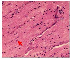
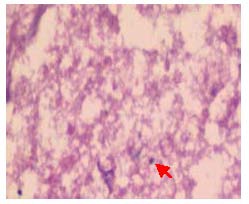
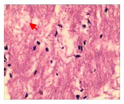
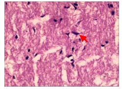
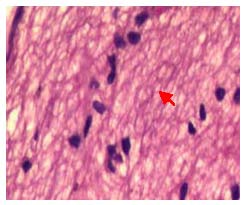
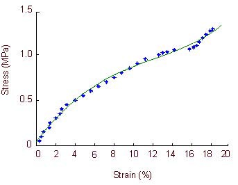
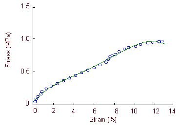
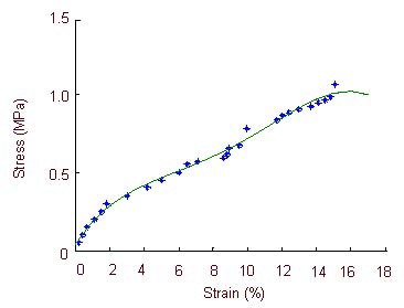
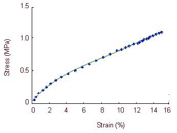
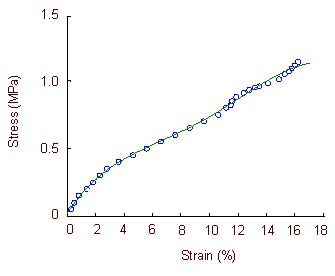
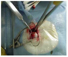
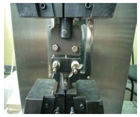
In this study, rabbit models of optic nerve injury were reproduced by the clamp method. After modeling, rabbit models were given one injection of 50 ng recombinant human ciliary neurotrophic factor into the vitreous body and/or intragastric injection of 4 g/kg compound light granules containing Radix Angelicae Sinensis and Raidix Paeoniae Alba at 4 days after modeling, once per day for 30 consecutive days. After administration, the animals were sacrificed and the intraorbital optic nerve was harvested. Hematoxylin-eosin staining revealed that the injured optic nerve was thinner and optic nerve fibers were irregular. After treatment with recombinant human ciliary neurotrophic factor, the arrangement of optic nerve fibers was disordered but they were not markedly thinner. After treatment with compound light granules, the arrangement of optic nerve fibers was slightly disordered and their structure was intact. After combined treatment with recombinant human ciliary neurotrophic factor and compound light granules, the arrangement of optic nerve fibers was slightly disordered and the degree of injury was less than after either treatment alone. Results of tensile mechanical testing of the optic nerve showed that the tensile elastic limit strain, elastic limit stress, maximum stress and maximum strain of the injured optic nerve were significantly lower than the normal optic nerve. After treatment with recombinant human ciliary neurotrophic factor and/or compound light granules, the tensile elastic limit strain, elastic limit stress, maximum stress and maximum strain of the injured optic nerve were significantly increased, especially after the combined treatment. These experimental findings indicate that compound light granules and ciliary neurotrophic factor can alleviate optic nerve injury at the histological and biochemical levels, and the combined treatment is more effective than either treatment alone.

Keywords: biomechanics; ciliary neurotrophic factor; compound light granules; mechanical characteristics; neural regeneration; optic nerve injury; retinal ganglial cells; strain; stress; tissue morphology; traditional Chinese medicine.


Conflict of interest statement

Conflicts of interest: None declared.

Figures

Figure 1
Figure 1
Morphology of a longitudinal section of the normal optic nerve in rabbits (hematoxylin-eosin staining, × 400). Fibers are densely arranged in parallel rows, with intact structure, uniform staining, and similar size and arrangement of glial cells (red arrow).
Figure 2
Figure 2
Pathological change in the longitudinal sections of the optic nerve in rabbits with optic nerve injury (hematoxylin-eosin staining, × 400). Optic nerve fiber structure became disorganized, nerve fibers and glial cells degenerated and became necrotic, and dissolved nuclei were occasionally observed (red arrow).
Figure 3
Figure 3
Pathological changes in the longitudinal sections of the optic nerve in rabbits after treatment with ciliary neurotrophic factor (hematoxylin-eosin staining, × 400). Optic nerve fibers arranged in disorder, the structure was visible, fiber bundles were not continuous, glial nuclei were increased, with some irregular nuclei, a small number of nuclei were dissolved and there were occasional nuclear fragments (red arrow).
Figure 4
Figure 4
Pathological changes in the longitudinal sections of the optic nerve in rabbits after treatment with compound light granules (hematoxylin-eosin staining, × 400). The structure of optic nerve fibers was visible; partial optic nerve fibers arranged irregularly, there was no thinning of the optic nerve, and glial nuclei were partially uneven (red arrow).
Figure 5
Figure 5
Pathological changes in the longitudinal sections of the optic nerve in rabbits after treatment with ciliary neurotrophic factor and compound light granules (hematoxylin-eosin staining, × 400). Optic nerve fibers arranged in disorder, fiber bundles were continuous (red arrow), glial nuclei were mostly regular, with a few irregular nuclei being observed.
Figure 6
Figure 6
Stress-strain curve of the optic nerve in the normal control group. In normal rabbits, the stress-strain curve is exponential when optic nerve strain is 0–3.8%, is directly proportional when optic nerve strain is 3.8–12.6%, and returns to an exponential relationship when optic strain is 12.6–15.8%. Large deformations occur in the specimens when the optic nerve strain is 15.8–18.2%, when the specimens lose their load-bearing capacity.
Figure 7
Figure 7
Stress-strain curve of the optic nerve in the model group. The stress-strain curve is exponential when optic nerve strain is 0–1.8% and 8.0–10.4%. Large deformations occur in the specimens when the optic nerve strain is 10.4–12.7%, when specimens lose their load-bearing capacity.
Figure 8
Figure 8
Stress-strain curve of the optic nerve in the ciliary neurotrophic factor group. The stress-strain curve is exponential when optic nerve strain is 0–2.2% and 9.45–11.7%. Large deformations occur in the specimens when the optic nerve strain is 11.7–15.1%, when specimens lose their load-bearing capacity.
Figure 9
Figure 9
Stress-strain curve of the optic nerve in the compound light granule group. The stress-strain curve is exponential when optic nerve strain is 0–2.5%, is directly proportional when optic nerve strain is 2.5–10.2%, and returns to an exponential relationship when optic strain is 10.2–12.2%. Large deformations occur in the specimens when the optic nerve strain is 12.2–15.6%, when specimens lose their load-bearing capacity.
Figure 10
Figure 10
Stress-strain curve of the optic nerve in the ciliary neurotrophic factor + compound light granule group. The stress-strain curve is exponential when optic nerve strain is –2.8%, and is directly proportional when optic nerve strain is 2.8–10.7%. Large deformations occur in the specimens when the optic nerve strain is 12.8–16.2%, when specimens lose their load-bearing capacity.
Figure 11
Figure 11
Establishment of optic nerve injury models. Freeing of the retrobulbar optic nerve and clamping of the optic nerve with a mosquito forcep.
Figure 12
Figure 12
Electronic universal testing machine for measuring the length and diameter of each optic nerve specimen.

References

    1. Sheng YM, Meng XL. Research progress of the optic nerve protective effect of Chinese medicine. Yiyao Daobao. 2007;26(10):1191–1193.
    1. Kamradt MC, Lu M, Werner ME, et al. The small heat shock protein alpha B-crystallin is a novel inhibitor of TRAIL-induced apoptosis that suppresses the activation of caspase-3. J Biol Chem. 2005;280(12):11059–11066. - PubMed
    1. Morozov V, Wawrousek EF. Caspase-dependent secondary lens fiber cell disintegration in alphaA-/alphaB-crystallin double-knockout mice. Development. 2006;133(5):813–821. - PubMed
    1. Munemasa Y, Kwong JM, Caprioli J, et al. The role of alphaA- and alphaB-crystallins in the survival of retinal ganglion cells after optic nerve axotomy. Invest Ophthalmol Vis Sci. 2009;50(8):3869–3875. - PubMed
    1. Ghosh JG, Houck SA, Doneanu CE, et al. The beta4-beta8 groove is an ATP-interactive site in the alpha crystallin core domain of the small heat shock protein, human alphaB crystallin. J Mol Biol. 2006;364(3):364–375. - PubMed