

Caveolin-1 regulates P2X7 receptor signaling in osteoblasts
- PMID: 25318104
- PMCID: PMC4281673
- DOI: 10.1152/ajpcell.00037.2014
Caveolin-1 regulates P2X7 receptor signaling in osteoblasts
Abstract
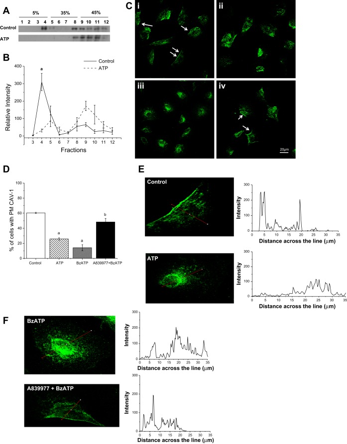
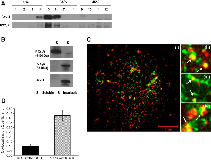
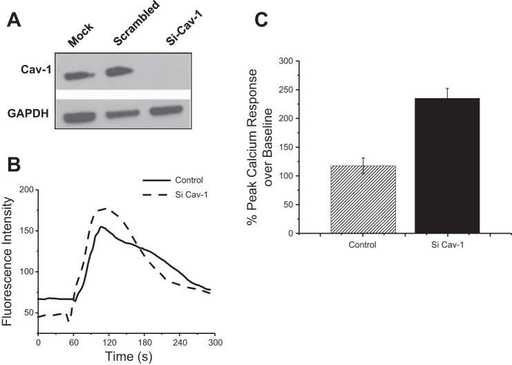
The synthesis of new bone in response to a novel applied mechanical load requires a complex series of cellular signaling events in osteoblasts and osteocytes. The activation of the purinergic receptor P2X(7)R is central to this mechanotransduction signaling cascade. Recently, P2X(7)R have been found to be associated with caveolae, a subset of lipid microdomains found in several cell types. Deletion of caveolin-1 (CAV1), the primary protein constituent of caveolae in osteoblasts, results in increased bone mass, leading us to hypothesize that the P2X(7)R is scaffolded to caveolae in osteoblasts. Thus, upon activation of the P2X(7)R, we postulate that caveolae are endocytosed, thereby modulating the downstream signal. Sucrose gradient fractionation of MC3T3-E1 preosteoblasts showed that CAV1 was translocated to the denser cytosolic fractions upon stimulation with ATP. Both ATP and the more specific P2X(7)R agonist 2'(3')-O-(4-benzoylbenzoyl)ATP (BzATP) induced endocytosis of CAV1, which was inhibited when MC3T3-E1 cells were pretreated with the specific P2X7R antagonist A-839977. The P2X7R cofractionated with CAV1, but, using superresolution structured illumination microscopy, we found only a subpopulation of P2X(7)R in these lipid microdomains on the membrane of MC3T3-E1 cells. Suppression of CAV1 enhanced the intracellular Ca(2+) response to BzATP, suggesting that caveolae regulate P2X(7)R signaling. This proposed mechanism is supported by increased mineralization in CAV1 knockdown MC3T3-E1 cells treated with BzATP. These data suggest that caveolae regulate P2X(7)R signaling upon activation by undergoing endocytosis and potentially carrying with it other signaling proteins, hence controlling the spatiotemporal signaling of P2X(7)R in osteoblasts.
Keywords: P2X7R; caveolin-1; endocytosis; osteoblasts; purinergic signaling.
Copyright © 2015 the American Physiological Society.
Figures

Similar articles
-
P2X₇-mediated calcium influx triggers a sustained, PI3K-dependent increase in metabolic acid production by osteoblast-like cells.Am J Physiol Endocrinol Metab. 2012 Mar 1;302(5):E561-75. doi: 10.1152/ajpendo.00209.2011. Epub 2011 Dec 20. Am J Physiol Endocrinol Metab. 2012. PMID: 22185840
-
PI3K/Akt/GSK-3β signal pathway is involved in P2X7 receptor-induced proliferation and EMT of colorectal cancer cells.Eur J Pharmacol. 2021 May 15;899:174041. doi: 10.1016/j.ejphar.2021.174041. Epub 2021 Mar 15. Eur J Pharmacol. 2021. PMID: 33737010
-
P2X(7) receptor antagonists display agonist-like effects on cell signaling proteins.Biochim Biophys Acta. 2011 May;1810(5):532-42. doi: 10.1016/j.bbagen.2011.03.009. Epub 2011 Mar 21. Biochim Biophys Acta. 2011. PMID: 21397667 Free PMC article.
-
Caveolar and non-Caveolar Caveolin-1 in ocular homeostasis and disease.Prog Retin Eye Res. 2022 Nov;91:101094. doi: 10.1016/j.preteyeres.2022.101094. Epub 2022 Jun 18. Prog Retin Eye Res. 2022. PMID: 35729002 Free PMC article. Review.
-
Roles of purinergic P2X7 receptor in glioma and microglia in brain tumors.Cancer Lett. 2017 Aug 28;402:93-99. doi: 10.1016/j.canlet.2017.05.004. Epub 2017 May 20. Cancer Lett. 2017. PMID: 28536012 Review.
Cited by
-
Methods for Studying Cholesterol-Dependent Regulation of P2X7 Receptors.Methods Mol Biol. 2022;2510:253-264. doi: 10.1007/978-1-0716-2384-8_13. Methods Mol Biol. 2022. PMID: 35776329
-
The mechanotransduction of MLO-Y4 cells is disrupted by the senescence-associated secretory phenotype of neighboring cells.J Cell Physiol. 2022 Apr;237(4):2249-2257. doi: 10.1002/jcp.30690. Epub 2022 Feb 1. J Cell Physiol. 2022. PMID: 35102547 Free PMC article.
-
Atheroprone flow activates inflammation via endothelial ATP-dependent P2X7-p38 signalling.Cardiovasc Res. 2018 Feb 1;114(2):324-335. doi: 10.1093/cvr/cvx213. Cardiovasc Res. 2018. PMID: 29126223 Free PMC article.
-
The Diminishing Returns of Mechanical Loading and Potential Mechanisms that Desensitize Osteocytes.Curr Osteoporos Rep. 2021 Aug;19(4):436-443. doi: 10.1007/s11914-021-00693-9. Epub 2021 Jul 3. Curr Osteoporos Rep. 2021. PMID: 34216359 Free PMC article. Review.
-
Caveolin-1 Regulates Parathyroid Hormone (PTH)-Related Protein (PTHrP) Actions on PTH Receptor Type 1 in Bone Cells.J Cell Physiol. 2025 Jul;240(7):e70067. doi: 10.1002/jcp.70067. J Cell Physiol. 2025. PMID: 40665637 Free PMC article.
References
-
- Alonso MA, Millan J. The role of lipid rafts in signalling and membrane trafficking in T lymphocytes. J Cell Sci 114: 3957–3965, 2001. - PubMed
-
- Barth K, Weinhold K, Guenther A, Young MT, Schnittler H, Kasper M. Caveolin-1 influences P2X7 receptor expression and localization in mouse lung alveolar epithelial cells. FEBS J 274: 3021–3033, 2007. - PubMed
-
- Bowler WB, Buckley KA, Gartland A, Hipskind RA, Bilbe G, Gallagher JA. Extracellular nucleotide signaling: a mechanism for integrating local and systemic responses in the activation of bone remodeling. Bone 28: 507–512, 2001. - PubMed
-
- Drab M, Verkade P, Elger M, Kasper M, Lohn M, Lauterbach B, Menne J, Lindschau C, Mende F, Luft FC, Schedl A, Haller H, Kurzchalia TV. Loss of caveolae, vascular dysfunction, and pulmonary defects in caveolin-1 gene-disrupted mice. Science 293: 2449–2452, 2001. - PubMed
Publication types
MeSH terms
Substances
Grants and funding
LinkOut - more resources
Full Text Sources
Other Literature Sources
Miscellaneous