. 2015 Jan;29(1):25-36.
doi: 10.1096/fj.14-253567. Epub 2014 Oct 15.

CD39 improves survival in microbial sepsis by attenuating systemic inflammation

Affiliations

CD39 improves survival in microbial sepsis by attenuating systemic inflammation

Balázs Csóka et al. FASEB J. 2015 Jan.

Abstract

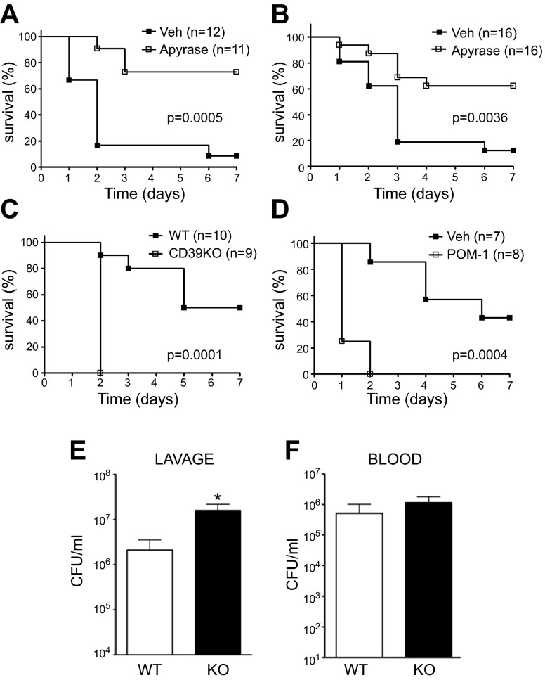
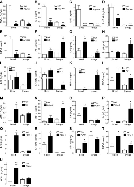
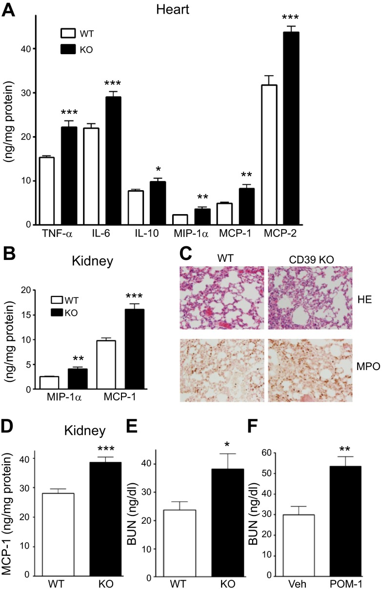
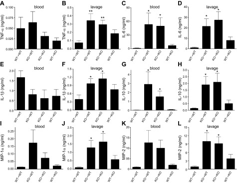
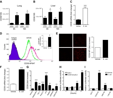
Sepsis remains the leading cause of morbidity and mortality in critically ill patients. Excessive inflammation is a major cause of organ failure and mortality in sepsis. Ectonucleoside triphosphate diphosphohydrolase 1, ENTPDase1 (CD39) is a cell surface nucleotide-metabolizing enzyme, which degrades the extracellular purines ATP and ADP, thereby regulating purinergic receptor signaling. Although the role of purinergic receptor signaling in regulating inflammation and sepsis has been addressed previously, the role of CD39 in regulating the host's response to sepsis is unknown. We found that the CD39 mimic apyrase (250 U/kg) decreased and knockout or pharmacologic blockade with sodium polyoxotungstate (5 mg/kg; IC50 ≈ 10 μM) of CD39 increased mortality of mice with polymicrobial sepsis induced by cecal ligation and puncture. CD39 decreased inflammation, organ damage, immune cell apoptosis, and bacterial load. Use of bone marrow chimeric mice revealed that CD39 expression on myeloid cells decreases inflammation in septic mice. CD39 expression is upregulated during sepsis in mice, as well as in both murine and human macrophages stimulated with Escherichia coli. Moreover, E. coli increases CD39 promoter activity in macrophages. Altogether, these data indicate CD39 as an evolutionarily conserved inducible protective pathway during sepsis. We propose CD39 as a novel therapeutic target in the management of sepsis.

Keywords: MIP; TNF; interleukin; kidney; lung.


Figures

Figure 1.
Figure 1.
CD39 improves survival in CLP-induced sepsis. Survival of vehicle- (Veh)- or apyrase-treated WT mice (A, B). Apyrase or its vehicle was administrated before CLP (A) or 6 h after CLP (B). Survival of CD39 KO and WT mice (C) and Veh- or POM-1–treated WT mice (D). Surviving mice were counted every day for 7 d after inducing polymicrobial sepsis by way of CLP. Numbers of bacterial CFUs in the peritoneum (E) or blood (F) of WT and CD39 KO animals 16 h after CLP. n = 8 mice/group. Data are presented as mean ± sem. *P < 0.05 and **P < 0.01 vs. WT littermates or sham-operated animals.
Figure 2.
Figure 2.
CD39 decreases cytokine and chemokine production in CLP-induced sepsis. Blood and peritoneal lavage levels of TNF-α (A), IL-6 (B), IL-10 (C), IL-12 p40 (D), and MIP-2 (E) in apyrase- or vehicle (Veh)-treated mice 16 h after CLP. Results are representative of 2 experiments, n = 10 mice/group. Blood and peritoneal lavage levels of TNF-α (F), IL-6 (G), IL-1β (H), IL-10 (I), IL-12 p40 (J), MIP-1α (K), MIP-2 (L), and MCP-1 (M) in CD39 KO and WT mice 16 h after CLP. n = 8 mice/group. Blood and peritoneal lavage levels of TNF-α (N), IL-6 (O), IL-1β (P), IL-10 (Q), IL-12 p40 (R), MIP-1α (S), MIP-2 (T), and MCP-1 (U) in POM-1– or Veh-treated mice 16 h after CLP. Results are representative of 2 experiments, n = 10 mice/group. Cytokine and chemokine levels were determined using ELISA. Data are presented as mean ± sem. *P < 0.05, **P < 0.01, and ***P < 0.001 vs. WT or Veh-treated mice.
Figure 3.
Figure 3.
CD39 protects against sepsis-induced organ injury and inflammation. A) Cytokine and chemokine levels in the heart of WT and CD39 KO animals 16 h after CLP, as determined by ELISA. B) Chemokine levels in the kidney of WT and CD39 KO animals 16 h after CLP. n = 8 mice/group. C) Tissue sections of lungs of septic CD39 KO and WT mice 6 h after CLP stained with hematoxylin and eosin (HE) (top) or with anti-myeloperoxidase antibody (bottom). A representative section is shown from slides from 8 WT and KO animals. The staining was visualized and images were acquired by BX-41 microscope and U-TV1x2 camera (Olympus) with UPlan FLN 20×/0.5 objective (f/1.1 1/240) at a final ×200 magnification, using cellSens Entry software (Olympus). For enhancing quality, TIF file images were further adjusted by Adobe Photoshop CS5 (Adobe Systems Incorporated, San Jose, CA, USA). Layers were duplicated with the mode changed to soft light. All images were processed with the same modifications. D) MCP-1 levels in the kidney of WT and CD39 KO mice 16 h after CLP. E and F) BUN levels in the plasma of CD39 KO vs. WT mice (E) and Veh- or POM-1–treated mice (F) 16 h after CLP. n = 8–10 mice/group. Data are presented as mean ± sem. *P < 0.05, **P < 0.01, and ***P < 0.001 vs. WT or Veh-treated mice.
Figure 4.
Figure 4.
CD39 deficiency on myeloid cells is responsible for the increased inflammatory response of CD39 KO mice. Blood and peritoneal lavage levels of TNF-α (A, B), IL-6 (C, D), IL-1β (E, F), IL-10 (G, H), MIP-1α (I, J), and MIP-2 (K, L) in WT→WT (CD39 WT bone marrow and WT parenchyma), KO→WT (CD39 KO bone marrow and WT parenchyma), KO→KO (CD39 KO bone marrow and KO parenchyma), and WT→KO (CD39 WT bone marrow and KO parenchyma) mice 16 h after CLP. n = 4, 8, 6, and 8 mice/group, respectively. Data are presented as mean ± sem. *P < 0.05 and **P < 0.01 vs. WT→WT mice.
Figure 5.
Figure 5.
Sepsis upregulates CD39 expression in vivo and in vitro. A, B) mRNA expression of CD39 in lung (A) and liver (B) of CLP- vs. sham-operated WT mice. n = 3–6 mice/group. C–F) Escherichia coli upregulates CD39 expression in murine (C–E) and human (F) THP-1 macrophages. RAW 264.7 macrophages were stimulated with heat-inactivated E. coli (15:1 bacteria/macrophage ratio) for 12 h. CD39 mRNA was detected using real-time PCR (C), and CD39 expression on the cell surface was determined using flow cytometry (D) or immune fluorescence (E). For the and immune fluorescent imaging (E), the fluorescence intensity was quantified using ImageJ software and was normalized to vehicle treatments. THP-1 macrophage-like cells were stimulated with heat-inactivated E. coli (15:1 bacteria/macrophage ratio) for 12 h. CD39 mRNA was detected using real-time PCR (F). G) TLR agonists increase CD39 gene expression in RAW264.7 macrophages. Cells were stimulated with 1 μg/ml PAM3CSK4, 1 μg/ml FSL-1, 10 μg/ml Poly(I:C), 10 μg/ml LPS, 10 μg/ml flagellin, 10 μg/ml ssRNA40, or 5 μM ODN1826 for 12 h. Murine CD39 mRNA was detected using real-time PCR. H) E. coli augments CD39 promoter activity in RAW 264.7 macrophages. RAW 264.7 cells were transiently transfected with full-length CD39 reporter (−1039/+65) or control pGL3 reporter plasmid. Transfected cells were treated with E. coli (1:15 macrophage/bacteria ratio) for 4, 8, or 16 h. Luciferase activities were determined using Dual-Luciferase Reporter Assay System. I) Role of promoter regions in the enhancing effect of E. coli on CD39 promoter activity in macrophages. RAW 264.7 cells were transfected with a series of CD39 promoter deletion mutants that were inserted in the pGL3 luciferase reporter vector. Transfected cells were stimulated with E. coli (1:15 macrophage/bacteria ratio) for 8 h. Luciferase activities are expressed as the mean activity and sem relative to the activity of the empty pGL3 vector after E. coli stimulation followed by normalization to protein concentration. Each result is representative of 3 experiments. Data are presented as mean ± sem. **P < 0.01 and ***P < 0.001 vs. vehicle or 0 h or pGL3.
Figure 6.
Figure 6.
Effect of ATP and AMP on IL-12 and IL-10 production by macrophages with inactivated CD39 or CD73. A, B) The effect of ATP on IL-12 p40 (A) and IL-10 (B) production by LPS-stimulated peritoneal macrophages in the absence (vehicle) or presence of POM-1. Peritoneal macrophages were stimulated with 10 μg/ml LPS or 300 μM ATP + LPS for 24 h in the absence of presence of POM-1, and then cytokine levels were measured from the supernatants. C, D) The effect of AMP on IL-12 p40 (A) and IL-10 (B) production by LPS-stimulated WT or CD73 peritoneal macrophages. CD73 KO or WT macrophages were stimulated with LPS or 300 μM AMP + LPS for 24 h. Results are representative of 3 experiments. Data are presented as mean ± sem. ***P < 0.001 vs. control (con, no LPS) group and #P < 0.05 or ###P < 0.001 vs. LPS.

References

    1. Van der Poll T., and Opal S. M. (2008) Host-pathogen interactions in sepsis. Lancet Infect. Dis. 8, 32–43 - PubMed
    1. Bone R. C., Balk R. A., Cerra F. B., Dellinger R. P., Fein A. M., Knaus W. A., Schein R. M., and Sibbald W. J.; ACCP/SCCM Consensus Conference Committee (2009) Definitions for sepsis and organ failure and guidelines for the use of innovative therapies in sepsis. The ACCP/SCCM Consensus Conference Committee. American College of Chest Physicians/Society of Critical Care Medicine. 1992. Chest 136(5, Suppl)e28. - PubMed
    1. Robertson C. M., and Coopersmith C. M. (2006) The systemic inflammatory response syndrome. Microbes Infect. 8, 1382–1389 - PubMed
    1. Stearns-Kurosawa D. J., Osuchowski M. F., Valentine C., Kurosawa S., and Remick D. G. (2011) The pathogenesis of sepsis. Annu. Rev. Pathol. 6, 19–48 - PMC - PubMed
    1. Xiao W., Mindrinos M. N., Seok J., Cuschieri J., Cuenca A. G., Gao H., Hayden D. L., Hennessy L., Moore E. E., Minei J. P., Bankey P. E., Johnson J. L., Sperry J., Nathens A. B., Billiar T. R., West M. A., Brownstein B. H., Mason P. H., Baker H. V., Finnerty C. C., Jeschke M. G., López M. C., Klein M. B., Gamelli R. L., Gibran N. S., Arnoldo B., Xu W., Zhang Y., Calvano S. E., McDonald-Smith G. P., Schoenfeld D. A., Storey J. D., Cobb J. P., Warren H. S., Moldawer L. L., Herndon D. N., Lowry S. F., Maier R. V., Davis R. W., and Tompkins R. G.; Inflammation and Host Response to Injury Large-Scale Collaborative Research Program (2011) A genomic storm in critically injured humans. J. Exp. Med. 208, 2581–2590 - PMC - PubMed

Publication types

MeSH terms