

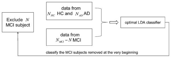
Baseline shape diffeomorphometry patterns of subcortical and ventricular structures in predicting conversion of mild cognitive impairment to Alzheimer's disease
- PMID: 25318546
- PMCID: PMC4474004
- DOI: 10.3233/JAD-141605
Baseline shape diffeomorphometry patterns of subcortical and ventricular structures in predicting conversion of mild cognitive impairment to Alzheimer's disease
Abstract
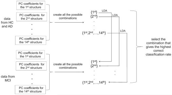
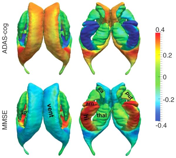
In this paper, we propose a novel predictor for the conversion from mild cognitive impairment (MCI) to Alzheimer's disease (AD). This predictor is based on the shape diffeomorphometry patterns of subcortical and ventricular structures (left and right amygdala, hippocampus, thalamus, caudate, putamen, globus pallidus, and lateral ventricle) of 607 baseline scans from the Alzheimer's Disease Neuroimaging Initiative database, including a total of 210 healthy control subjects, 222 MCI subjects, and 175 AD subjects. The optimal predictor is obtained via a feature selection procedure applied to all of the 14 sets of shape features via linear discriminant analysis, resulting in a combination of the shape diffeomorphometry patterns of the left hippocampus, the left lateral ventricle, the right thalamus, the right caudate, and the bilateral putamen. Via 10-fold cross-validation, we substantiate our method by successfully differentiating 77.04% (104/135) of the MCI subjects who converted to AD within 36 months and 71.26% (62/87) of the non-converters. To be specific, for the MCI-converters, we are capable of correctly predicting 82.35% (14/17) of subjects converting in 6 months, 77.5% (31/40) of subjects converting in 12 months, 74.07% (20/27) of subjects converting in 18 months, 78.13% (25/32) of subjects converting in 24 months, and 73.68% (14/19) of subject converting in 36 months. Statistically significant correlation maps were observed between the shape diffeomorphometry features of each of the 14 structures, especially the bilateral amygdala, hippocampus, lateral ventricle, and two neuropsychological test scores--the Alzheimer's Disease Assessment Scale-Cognitive Behavior Section and the Mini-Mental State Examination.
Keywords: Alzheimer's disease; lateral ventricles; linear discriminant analysis; mild cognitive impairment; prediction; principal component analysis; shape diffeomorphometry; subcortical structures.
Figures
References
-
- McKhann G, Drachman D, Folstein M, Katzman R, Price D, Stadlan EM. Clinical diagnosis of Alzheimer’s disease: Report of the NINCDS-ADRDA Work Group∗ under the auspices of Department of Health and Human Services Task Force on Alzheimer’s Disease. Neurology. 1984;34:939–939. - PubMed
-
- Gauthier S, Reisberg B, Zaudig M, Petersen RC, Ritchie K, Broich K, Belleville S, Brodaty H, Bennett D, Chertkow H, Cummings JL, de Leon M, Feldman H, Ganguli M, Hampel H, Scheltens P, Tierney MC, Whitehouse P, Winblad B, International Psychogeriatric Association. Expert Conference on mild cognitive impairment Mild cognitive impairment. Lancet. 2006;367:1262–1270. - PubMed
-
- Morris JC, Storandt M, Miller JP, McKeel DW, Price JL, Rubin EH, Berg L. Mild cognitive impairment represents early-stage Alzheimer disease. Arch Neurol. 2001;58:397–405. - PubMed
-
- Ramani A, Jensen JH, Helpern JA. Quantitative MR imaging in Alzheimer disease. Radiology. 2006;241:26–44. - PubMed
