

Thick ascending limb of the loop of Henle
- PMID: 25318757
- PMCID: PMC4220766
- DOI: 10.2215/CJN.04480413
Thick ascending limb of the loop of Henle
Abstract
The thick ascending limb occupies a central anatomic and functional position in human renal physiology, with critical roles in the defense of the extracellular fluid volume, the urinary concentrating mechanism, calcium and magnesium homeostasis, bicarbonate and ammonium homeostasis, and urinary protein composition. The last decade has witnessed tremendous progress in the understanding of the molecular physiology and pathophysiology of this nephron segment. These advances are the subject of this review, with emphasis on particularly recent developments.
Keywords: Na transport; acidosis; calcium receptor; renal physiology; water transport.
Copyright © 2014 by the American Society of Nephrology.
Figures
Similar articles
-
Ion transport mechanisms in thick ascending limb of Henle's loop of mammalian nephron.Physiol Rev. 1985 Jul;65(3):760-97. doi: 10.1152/physrev.1985.65.3.760. Physiol Rev. 1985. PMID: 2409564 Review. No abstract available.
-
A survey of transport properties of the thick ascending limb.Semin Nephrol. 1991 Mar;11(2):236-47. Semin Nephrol. 1991. PMID: 2034927 Review. No abstract available.
-
A possible catalytic role for NH4+ in Na+ reabsorption across the thick ascending limb.Am J Physiol Renal Physiol. 2010 Mar;298(3):F510-1. doi: 10.1152/ajprenal.00678.2009. Epub 2009 Dec 9. Am J Physiol Renal Physiol. 2010. PMID: 20007343 Free PMC article. No abstract available.
-
Thick ascending limb of Henle's loop.Kidney Int. 1982 Nov;22(5):454-64. doi: 10.1038/ki.1982.198. Kidney Int. 1982. PMID: 6296522 Review.
-
Heterogeneity of tight junctions in the thick ascending limb.Ann N Y Acad Sci. 2017 Oct;1405(1):5-15. doi: 10.1111/nyas.13400. Epub 2017 Jun 19. Ann N Y Acad Sci. 2017. PMID: 28628195 Review.
Cited by
-
The "3Ds" of Growing Kidney Organoids: Advances in Nephron Development, Disease Modeling, and Drug Screening.Cells. 2023 Feb 8;12(4):549. doi: 10.3390/cells12040549. Cells. 2023. PMID: 36831216 Free PMC article. Review.
-
The importance of the thick ascending limb of Henle's loop in renal physiology and pathophysiology.Int J Nephrol Renovasc Dis. 2018 Feb 15;11:81-92. doi: 10.2147/IJNRD.S154000. eCollection 2018. Int J Nephrol Renovasc Dis. 2018. PMID: 29497325 Free PMC article. Review.
-
Associations of CKD risk factors and longitudinal changes in urine biomarkers of kidney tubules among women living with HIV.BMC Nephrol. 2021 Aug 30;22(1):296. doi: 10.1186/s12882-021-02508-6. BMC Nephrol. 2021. PMID: 34461840 Free PMC article.
-
The Role of Renal Medullary Bilirubin and Biliverdin Reductase in Angiotensin II-Dependent Hypertension.Am J Hypertens. 2025 Mar 17;38(4):240-247. doi: 10.1093/ajh/hpae150. Am J Hypertens. 2025. PMID: 39656666
-
Flow-dependent transport processes 2024: filtration, absorption, and secretion.Am J Physiol Renal Physiol. 2025 May 1;328(5):F627-F637. doi: 10.1152/ajprenal.00303.2024. Epub 2025 Mar 17. Am J Physiol Renal Physiol. 2025. PMID: 40094947 Free PMC article.
References
-
- Rampoldi L, Scolari F, Amoroso A, Ghiggeri G, Devuyst O: The rediscovery of uromodulin (Tamm-Horsfall protein): From tubulointerstitial nephropathy to chronic kidney disease. Kidney Int 80: 338–347, 2011 - PubMed
-
- Nielsen S, Pallone T, Smith BL, Christensen EI, Agre P, Maunsbach AB: Aquaporin-1 water channels in short and long loop descending thin limbs and in descending vasa recta in rat kidney. Am J Physiol 268: F1023–F1037, 1995 - PubMed
-
- Tsuruoka S, Koseki C, Muto S, Tabei K, Imai M: Axial heterogeneity of potassium transport across hamster thick ascending limb of Henle’s loop. Am J Physiol 267: F121–F129, 1994 - PubMed
-
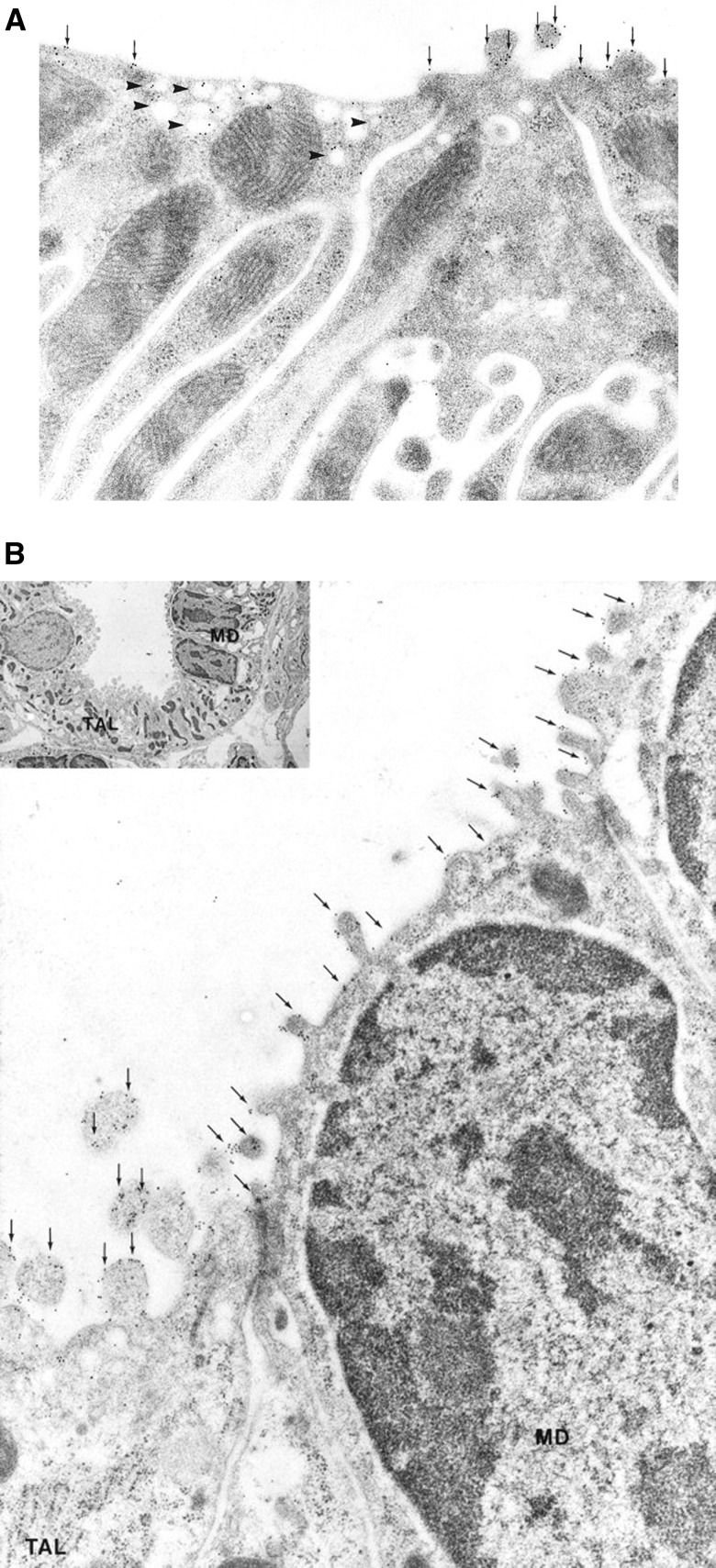
- Nielsen S, Maunsbach AB, Ecelbarger CA, Knepper MA: Ultrastructural localization of Na-K-2Cl cotransporter in thick ascending limb and macula densa of rat kidney. Am J Physiol 275: F885–F893, 1998 - PubMed
Publication types
MeSH terms
Substances
Grants and funding
LinkOut - more resources
Full Text Sources
Other Literature Sources
Medical
