. 2014 Oct 28;111(43):E4623-31.
doi: 10.1073/pnas.1418402111. Epub 2014 Oct 15.

Dynamic architecture of a protein kinase

Affiliations

Dynamic architecture of a protein kinase

Christopher L McClendon et al. Proc Natl Acad Sci U S A. .

Erratum in

  • Proc Natl Acad Sci U S A. 2014 Nov 25;111(47):16973

Abstract

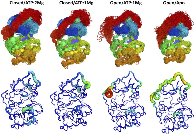
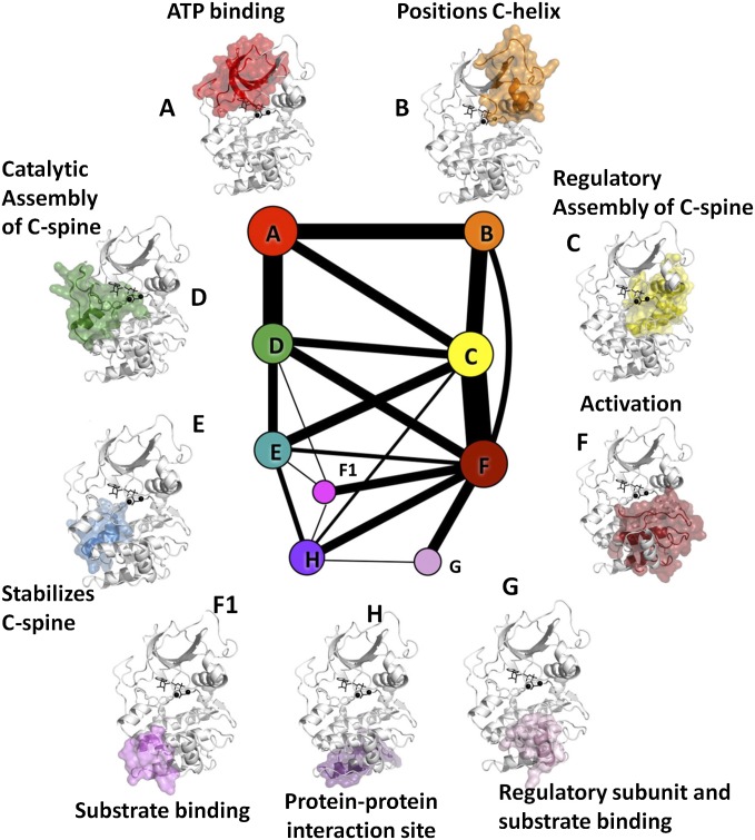
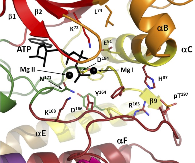
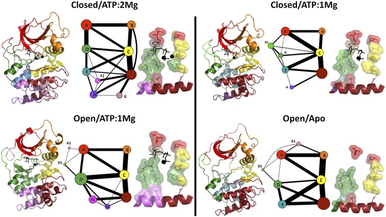
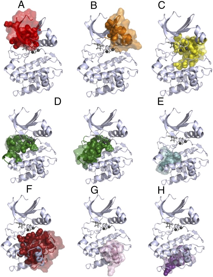
Protein kinases are dynamically regulated signaling proteins that act as switches in the cell by phosphorylating target proteins. To establish a framework for analyzing linkages between structure, function, dynamics, and allostery in protein kinases, we carried out multiple microsecond-scale molecular-dynamics simulations of protein kinase A (PKA), an exemplar active kinase. We identified residue-residue correlated motions based on the concept of mutual information and used the Girvan-Newman method to partition PKA into structurally contiguous "communities." Most of these communities included 40-60 residues and were associated with a particular protein kinase function or a regulatory mechanism, and well-known motifs based on sequence and secondary structure were often split into different communities. The observed community maps were sensitive to the presence of different ligands and provide a new framework for interpreting long-distance allosteric coupling. Communication between different communities was also in agreement with the previously defined architecture of the protein kinase core based on the "hydrophobic spine" network. This finding gives us confidence in suggesting that community analyses can be used for other protein kinases and will provide an efficient tool for structural biologists. The communities also allow us to think about allosteric consequences of mutations that are linked to disease.

Keywords: community analysis; molecular dynamics; phosphorylation.


Conflict of interest statement

Conflict of interest statement: M.K.G has an equity interest in, and is a cofounder and scientific advisor of, VeraChem LLC.

Figures

Fig. 1.
Fig. 1.
MD simulations of PKA in different ligand and conformational states, as noted, show that the conformation and flexibility of the G-helix and C-terminal tail are particularly sensitive to active site ligands. (Upper) Superimposed MD trajectory snapshots of the PKA backbone illustrate the conformational space sampled in the simulations, including differences among the various ligand and conformational states. The backbone of PKA is colored from blue to red according to position in the primary sequence from the N terminus to the C terminus, so that the C-tail is red. (Lower) Backbone root-mean-squared position fluctuations highlight the most dynamic regions of PKA in various liganded and conformational states. The largest differences in flexibility are seen in the C-terminal tail and the G-helix.
Fig. 2.
Fig. 2.
Community map of PKA, for the closed conformation with ATP and two Mg bound. The size of each node represents the number of members (residue backbones and side-chains) of each community, and the edge weights are proportional to the thickness of the lines. The full kinase is shown in white, the communities are highlighted in red, and ligands are shown in black. The legend annotates each community by its function.
Fig. 3.
Fig. 3.
Multiple communities converge around the ATP-binding and catalytic site. ComD provides catalytically critical N171, which coordinates Mg II and D166, a proton acceptor for the substrate’s –OH group in catalysis.
Fig. 4.
Fig. 4.
The N-lobe is subdivided into three communities that are largely preserved across different ligand/conformational states. (A) In ComA, we show a set of bridging residues’ side-chains in surface representation. (B) In ComB, a chiefly hydrophobic set of residues connects the hydrophobic motif (F347, F350) to the αC-helix and Gly-rich loop. (C) Previously identified subdivisions of the C-terminal tail segregate into different communities, and the entire tail connects communities A, B, D, and E.
Fig. 5.
Fig. 5.
ComC structurally connects the N-lobe to the C-lobe through the R-spine, DFG-motif, and αA-helix. (A) In ComC, surfaces show the R-spine and a set of cation–π interactions that connect the top of the αE-helix, the activation loop, and the αA- and αC-helices. (B) The αC-β4 region connects communities A, C, D, and E, providing a link between the R spine, ATP, and the C-lobe.
Fig. 6.
Fig. 6.
ComD and ComF are C-lobe communities that connect the C-lobe to the N-lobe. ComD (green) encompasses the αD-helix and part of the catalytic loop, coordinating ATP and Mg from below, and includes the majority of the C-spine (surface). Other C-spine residues are colored by their community. ComF (dark red) involves a large section of the C-lobe and includes a set of chiefly polar residues (surfaces) that nucleate around the activation loop phosphate (pT197, yellow) and connect the C-helix to the activation loop, F-helix, and finally AGC insert, involving a conserved salt bridge between E208 and R280. The activation segment is split across two communities and structurally links the C-helix to ATP/Mg, the phosphorylation site, and to the bottom of the C-lobe through the conserved salt bridge between E208 and R280. Additionally, Y204 connects this segment to E230, a critical substrate-binding element in Com-H.
Fig. 7.
Fig. 7.
Community maps of PKA are similar across different ligand and conformational states and reveal conserved cores. For each of the four ligand and conformational states, the community map is shown with node sizes proportional to the relative number of community members and edge weights proportional to the total mutual information between the communities. To the left of each map, the structure of PKA is colored by community. To the right of each community map, the R-spine and C-spine are shown and colored by community; ATP is shown in sticks, and the C-spine and R-spine are shown in surface representations.
Fig. 8.
Fig. 8.
Community maps (A–H) of PKA in different conformational and ligand states reveal cores (surfaces), members that remain in the same community with each other. These cores contain many of the community-forming residues (spheres). In ComD, because there are two conserved cores, community-forming residues that belong to the other core are not shown.

References

    1. Johnson DA, Akamine P, Radzio-Andzelm E, Madhusudan M, Taylor SS. Dynamics of cAMP-dependent protein kinase. Chem Rev. 2001;101(8):2243–2270. - PubMed
    1. Huse M, Kuriyan J. The conformational plasticity of protein kinases. Cell. 2002;109(3):275–282. - PubMed
    1. Taylor SS, Keshwani MM, Steichen JM, Kornev AP. Evolution of the eukaryotic protein kinases as dynamic molecular switches. Philos Trans R Soc Lond B Biol Sci. 2012;367(1602):2517–2528. - PMC - PubMed
    1. Shaw AS, Kornev AP, Hu J, Ahuja LG, Taylor SS. Kinases and pseudokinases: Lessons from RAF. Mol Cell Biol. 2014;34(9):1538–1546. - PMC - PubMed
    1. Hanks SK, Quinn AM, Hunter T. The protein kinase family: Conserved features and deduced phylogeny of the catalytic domains. Science. 1988;241(4861):42–52. - PubMed

Publication types