. 2014 Oct 28;111(43):15402-7.
doi: 10.1073/pnas.1320644111. Epub 2014 Oct 15.

DNA supercoiling: a regulatory signal for the λ repressor

Affiliations

DNA supercoiling: a regulatory signal for the λ repressor

Yue Ding et al. Proc Natl Acad Sci U S A. .

Abstract

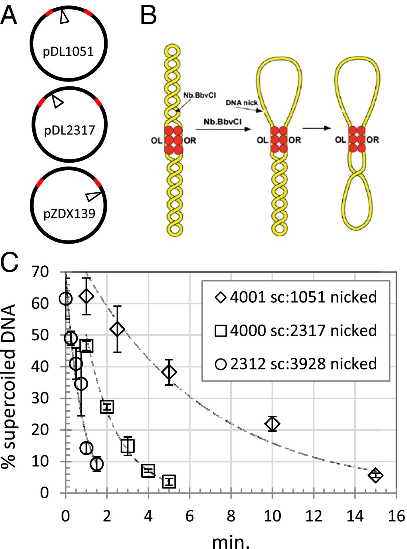
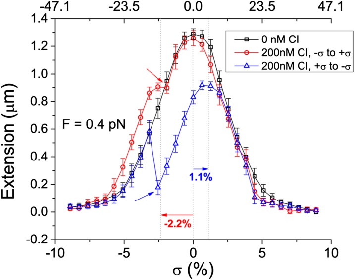
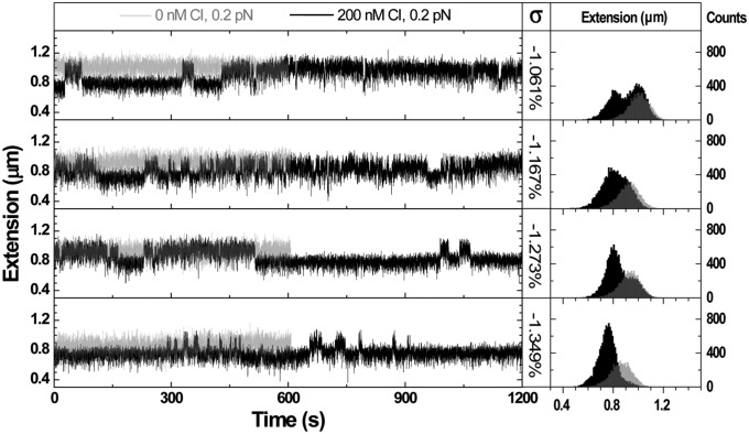
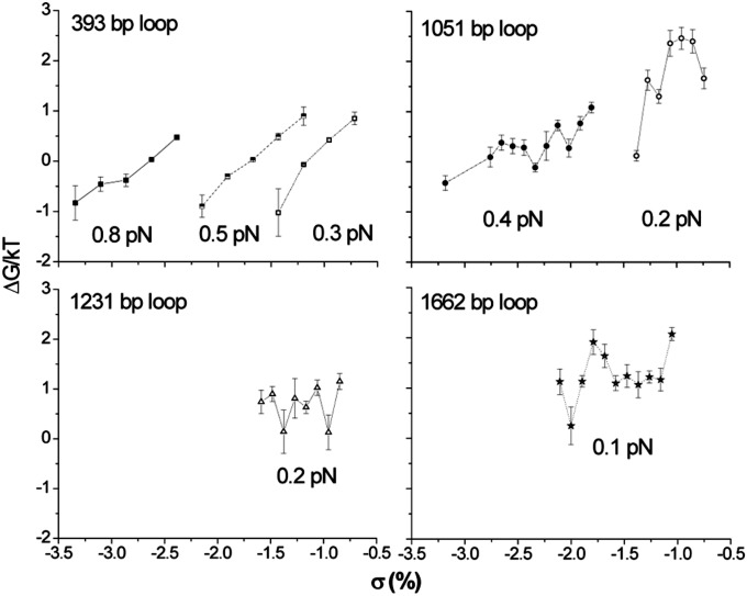
Topoisomerases, polymerases, and the chirality introduced by the binding of histones or nucleoid-associated proteins affect DNA supercoiling in vivo. However, supercoiling is not just a by-product of DNA metabolism. Supercoiling is an indicator of cell health, it modifies the accessibility of chromatin, and coordinates the transcription of genes. This suggests that regulatory, protein-mediated loops in DNA may sense supercoiling of the genome in which they are embedded. The λ repressor (CI) maintains the quiescent (lysogenic) transcriptome of bacteriophage λ in infected Escherichia coli. CI-mediated looping prevents overexpression of the repressor protein to preserve sensitivity to conditions that trigger virulence (lysis). Experiments were performed to assess how well the CI-mediated DNA loop traps superhelicity and determine whether supercoiling enhances CI-mediated DNA looping. CI oligomers partitioned plasmids into topological domains and prevented the passage of supercoiling between them. Furthermore, in single DNA molecules stretched and twisted with magnetic tweezers, levels of superhelical density confined in CI-mediated DNA loops ranged from -15% or +11%. Finally, in DNA under tensions that may occur in vivo, supercoiling lowered the free energy of loop formation and was essential for DNA looping. Supercoiling-enhanced looping can influence the maintenance of lysogeny in the λ repressor system; it can encode sensitivity to the energy level of the cell and creates independent topological domains of distinct superhelical density.

Keywords: DNA looping; DNA supercoiling; bacteriophage λ repressor; magnetic tweezer; transcription.


Conflict of interest statement

The authors declare no conflict of interest.

Figures

Fig. 1.
Fig. 1.
Supercoil diffusion through CI barriers. (A) The location of a nicking site for the enzyme Nb.BbvCI is shown relative to the OR and OL operators in plasmids pDL1051, pDL2317, and pZDX139. Red segments represent λ operators OL1–3 and OR1–3. (B) A topological barrier experiment with CI (red circles). Upon binding to OL and OR operators, the loop securing CI functions as a barrier that blocks supercoil diffusion. A DNA nick introduced by the endonuclease Nb.BbvCI quickly relaxes superhelicity within the upper loop. Superhelicity in the bottom loop slowly diffuses through the CI-mediated topological barrier where it then dissipates quickly. (C) Supercoiling slowly dissipated from supercoiled domains of plasmids partitioned by a CI-mediated loop. DNA-nicking assays were as described in Materials and Methods (Figs. S1 and S2). The percentage of remaining supercoiled DNA was plotted as a function of the incubation time. Fitting with an exponential decay yielded time constants of 0.74, 1.5, or 6.1 min for partitions of (supercoiled/nicked) 2,312:3,928, 4,000:2,317, or 4,001:1,051 bp, respectively.
Fig. 2.
Fig. 2.
Representative extension vs. twist curves showing shifted and reduced maxima upon loop formation. Under 0.4 pN of tension in the absence of CI protein (black), a tether with a 1,051-bp loop formed plectonemes that reduced extension for both positive and negative twist. With 200 nM CI protein (red and blue), as the twist was relaxed from −10% (red) or 10% (blue) superhelical density levels (bottom axis), reduced maxima were observed with shifts (horizontal arrows) that indicated the change in superhelical density of the looped tether (top axis). The superhelical density, σ, of these loops was –0.115 (red) or 0.057 (blue). Upon rupture of the loops (inclined arrows), the tether extension suddenly returned to unlooped values. Series of points are connected by segments to facilitate understanding. Error bars represent the SDs of the length measurements.
Fig. 3.
Fig. 3.
Negative superhelical density favors DNA loop formation. Representative recordings of extension vs. time (Left) and respective extension histograms (Right) for single DNA tethers under 0.2 pN tension, show the formation and rupture of 1,051-bp loops at different supercoiling levels. In the absence of CI protein, the DNA extension was constant appearing as a single peak on the respective extension histogram. The peak corresponding to the unlooped tether shifted slightly toward shorter values as negative superhelical density increased. In the presence of 200 nM CI, the extension of the tether intermittently shifted between looped and unlooped configurations creating telegraphic signals that gave rise to bimodal histograms. An additional −0.3% of twist was enough to shift the DNA from nearly evenly splitting time in looped and unlooped states to remaining almost always looped.
Fig. 4.
Fig. 4.
The calculated free energy (∆G) for the formation of loops under different tensions and superhelical density levels based on the lifetimes of looped and unlooped states. For identical loops under identical tension, more negative superhelical density decreased ∆G. This trend was clearer for smaller loops (393 and 1,051 bp) than for bigger ones (1,231 and 1,662 bp).

References

    1. Filipski J, Leblanc J, Youdale T, Sikorska M, Walker PR. Periodicity of DNA folding in higher order chromatin structures. EMBO J. 1990;9(4):1319–1327. - PMC - PubMed
    1. Gibcus JH, Dekker J. The hierarchy of the 3D genome. Mol Cell. 2013;49(5):773–782. - PMC - PubMed
    1. Naughton C, et al. Transcription forms and remodels supercoiling domains unfolding large-scale chromatin structures. Nat Struct Mol Biol. 2013;20(3):387–395. - PMC - PubMed
    1. Rovinskiy N, Agbleke AA, Chesnokova O, Pang Z, Higgins NP. Rates of gyrase supercoiling and transcription elongation control supercoil density in a bacterial chromosome. PLoS Genet. 2012;8(8):e1002845. - PMC - PubMed
    1. Postow L, Hardy CD, Arsuaga J, Cozzarelli NR. Topological domain structure of the Escherichia coli chromosome. Genes Dev. 2004;18(14):1766–1779. - PMC - PubMed

Publication types

Substances