Observational Study
. 2014 Oct;93(18):e116.
doi: 10.1097/MD.0000000000000116.

Effect of switching therapy to pegaptanib in eyes with the persistent cases of exudative age-related macular degeneration

Affiliations
Observational Study

Effect of switching therapy to pegaptanib in eyes with the persistent cases of exudative age-related macular degeneration

Chieko Shiragami et al. Medicine (Baltimore). 2014 Oct.

Abstract

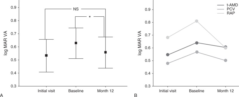
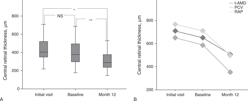
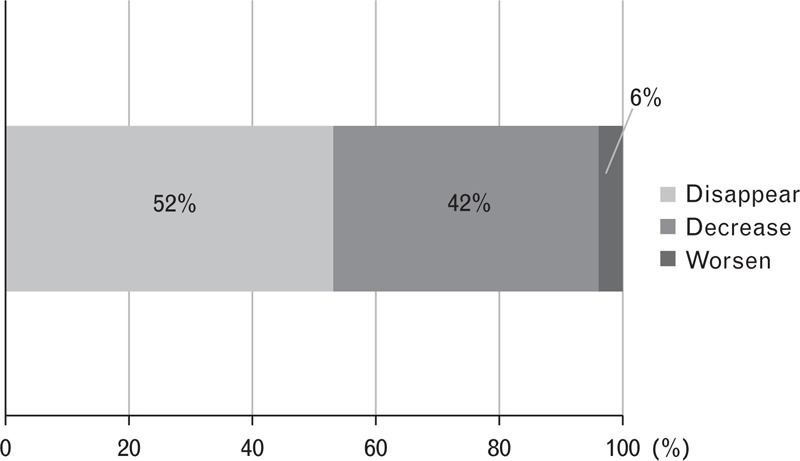
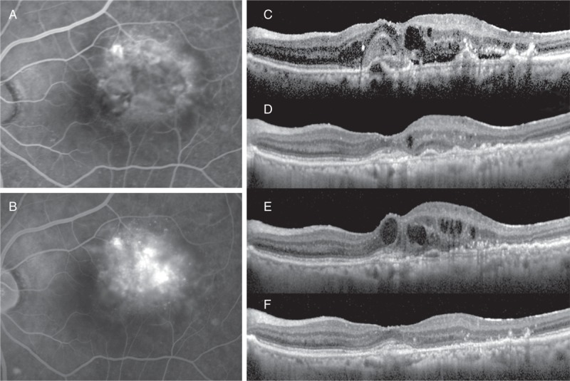
Purpose of this study was to evaluate the efficacy of switching to pegaptanib monotherapy for persistent cases of exudative age-related macular degeneration (AMD).Out of 296 eyes of 296 patients treated with ranibizumab or ranibizumab combined with photodynamic therapy (PDT), 50 eyes of 50 AMD patients were found to be resistant to these treatments. Over a 12-month period, intravitreal pegaptanib (IVP) 0.3 mg was administered at intervals of 6 weeks until the exudation disappeared prospectively. All patients were examined with the following tests: best-corrected visual acuity (BCVA) and central retinal thickness (CRT), determined at the initial visit, before the first IVP (baseline), and at 12 months. The factors responsible for achieving dry macula with IVP were examined statistically.The rate of persistent cases with intravitreal ranibizumab (IVR) and/or PDT was 17.0%. The mean number of IVPs administered was 5.4 (range, 2-9). Logarithm of the minimal angle of resolution BCVA at 12 months was stable or improved by ≥ 0.3 in 49 eyes (98.0%), with a significant improvement noted between the baseline and final BCVA (P=0.01, paired t test). The CRT (mean ± standard deviation) was 446.9 ± 150.6 µm at the initial visit, 414.5 ± 146.5 µm at baseline, and 318.7 ± 99.0 µm at 12 months. There was a significant decrease in the mean CRT between the measurements at baseline and at 12 months after the first IVP (P=0.002, Bonferroni correction). At 12 months, the exudative change was completely resolved in 27 eyes (54.0%) and reduced in 21 eyes (42.0%). The number of previous IVR treatments was significantly correlated with dry macula at 12 months.After switching therapy to pegaptanib in persistent cases of AMD, most patients maintained or improved their BCVA and exhibited a positive treatment response at 12 months.


Conflict of interest statement

The authors have no funding to disclose.

The authors have no proprietary interest in any aspect of this study.

Figures

FIGURE 1
FIGURE 1
(A) Mean change in logMAR VA (whole type of AMD cases). This plot graph shows the changes in the mean BCVA in logMAR units at the initial visit, baseline, and at 12 months. The error bar values indicate the standard deviation. The logMAR BCVA (mean ± standard deviation) was 0.53 ± 0.44 at the initial visit, 0.63 ± 0.41 at baseline, and 0.56 ± 0.42 at 12 months. A significant improvement was seen between the baseline and the BCVA at the last visit (∗P = 0.01, paired t test). (B) Mean change in logMAR VA (each type of AMD cases).   P value indicates the statistical correlation of logMAR BCVA between baseline and 12 months (paired t test). AMD = age-related macular degeneration, BCVA = best-corrected visual acuity, logMAR = logarithm of the minimal angle of resolution, NS = not significant, PCV = polypoidal choroidal vasculopathy, RAP = retinal angiomatous proliferation, t-AMD = typical AMD, VA = visual acuity.
FIGURE 2
FIGURE 2
(A) Mean change in CRT (whole type of AMD cases). Box plots show the CRT at the initial visit, baseline, and at 12 months. The CRT (mean ± standard deviation) was 446.9 ± 150.6 μm at the initial visit, 414.5 ± 146.5 μm at baseline, and 317.1 ± 99.1 μm at 12 months. A significant improvement in the CRT was seen between the initial visit and the measurements at 12 months (∗P < 0.001) and between the baseline and the measurements at 12 months (∗∗P = 0.002) (Bonferroni correction). (B) Mean change in CRT (each type of AMD cases).   P value indicates the statistical correlation of CRT between baseline and 12 months (paired t test). AMD = age-related macular degeneration, CRT = central retinal thickness, NS = not significant, PCV = polypoidal choroidal vasculopathy, RAP = retinal angiomatous proliferation, t-AMD = typical AMD.
FIGURE 3
FIGURE 3
The bar graph shows the change of the exudative lesion from the baseline to 12 months for 50 eyes with AMD, switched from ranibizumab and/or combined PDT to pegaptanib monotherapy. The exudative lesion disappeared completely in 52%, decreased in 42%, and worsened in 6%. AMD = age-related macular degeneration, PDT = photodynamic therapy.
FIGURE 4
FIGURE 4
In case 1, the patient was a 74-year-old man with PCV who was resistant to the treatment with ranibizumab-combined PDT. Subfoveal polypoidal lesion and vascular network was showed on early phase ICGA (A). The OCT revealed PED and SRD (B). Although the exudative lesion disappeared after 3 IVR-combined PDT (C), the PED and SRD recurred after 4 additional IVR (D). After that, ranibizumab was switched to pegaptanib; there was complete resolution of the PED and SRD (E). ICGA = indocyanine green angiography, IVR = intravitreal ranibizumab, OCT = optical coherence tomography, PCV = polypoidal choroidal vasculopathy, PDT = photodynamic therapy, PED = pigment epithelial detachment, SRD = serous retinal detachment.
FIGURE 5
FIGURE 5
In case 2, the patient was a 90-year-old woman with t-AMD who was resistant to the treatment with ranibizumab monotherapy. The FA showed minimally classic CNV (A, early phase; B, late phase), and OCT revealed the presence of SRD, small PED, and CME (C). The exudative lesion almost resolved after 9 IVRs at 12 months (D). At 3 months after the last IVR, because there was exacerbation of the CME (E), ranibizumab was switched to pegaptanib. At 12 months after baseline, there was complete resolution of the CME (F). AMD = age-related macular degeneration, CNV = choroidal neovascularization, CME = cystoid macular edema, CRT = central retinal thickness, FA = fluorescein angiography, IVR = intravitreal ranibizumab, OCT = optical coherence tomography, RD = subretinal detachment, PED = pigment epithelial detachment, SRD = serous retinal detachment, t-AMD = typical AMD.

Similar articles

Cited by

References

    1. Apte RS, Modi M, Masonson H, et al. ; Macugen AMD Study Group. Pegaptanib 1-year systemic safety results from a safety-pharmacokinetic trial in patients with neovascular age-related macular degeneration. Ophthalmology. 2007;114:1702–1712. - PubMed
    1. Takeda AL, Colquitt J, Clegg AJ, et al. Pegaptanib and ranibizumab for neovascular age-related macular degeneration: a systematic review. Br J Ophthalmol. 2007;91:1177–1182. - PMC - PubMed
    1. Rosenfeld PJ, Brown DM, Heier JS, et al. Ranibizumab for neovascular age-related macular degeneration. N Engl J Med. 2006;355:1419–1431. - PubMed
    1. Boyer DS, Antoszyk AN, Awh CC, et al. ; MARINA Study Group. Subgroup analysis of the MARINA study of ranibizumab in neovascular age-related macular degeneration. Ophthalmology. 2007;114:246–252. - PubMed
    1. The CATT Research Group. Ranibizumab and Bevacizumab for neovascular age-related macular degeneration. N Engl J Med. 2011;1–13. - PMC - PubMed

Publication types

MeSH terms