Randomized Controlled Trial
. 2014 Oct 15;34(42):13924-34.
doi: 10.1523/JNEUROSCI.1648-14.2014.

Selective activation of ipsilateral motor pathways in intact humans

Affiliations
Randomized Controlled Trial

Selective activation of ipsilateral motor pathways in intact humans

Toshiki Tazoe et al. J Neurosci. .

Abstract

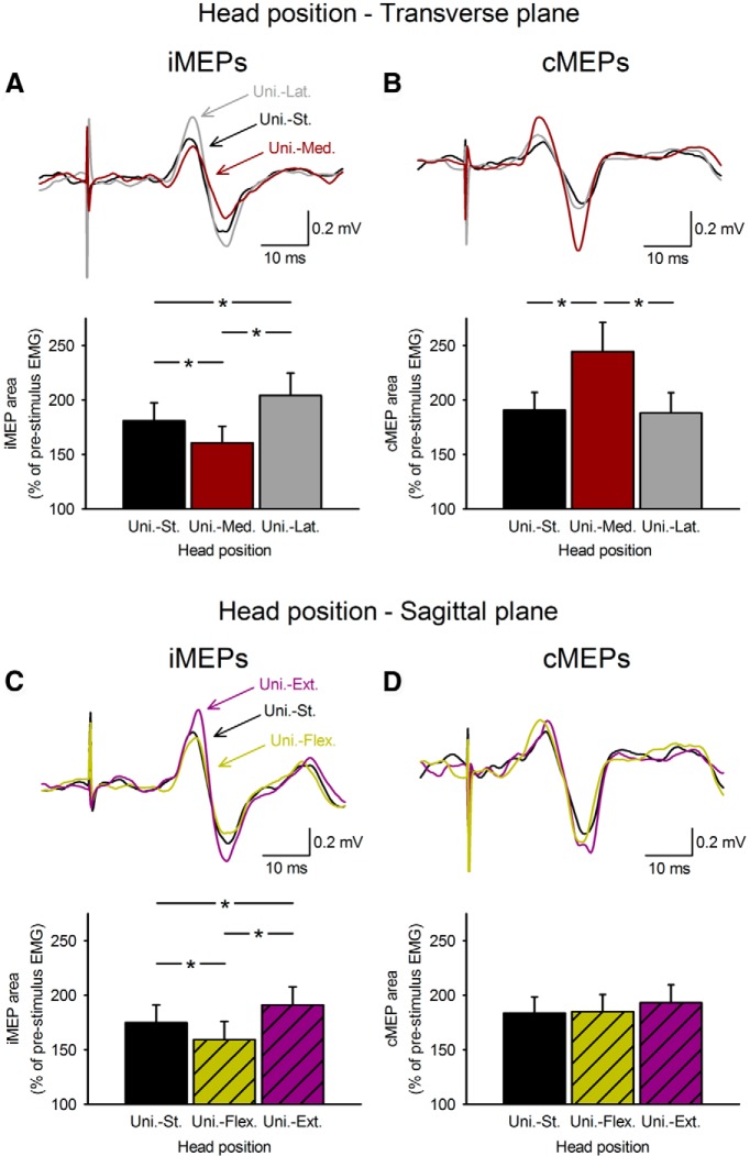
It has been proposed that ipsilateral motor pathways play a role in the control of ipsilateral movements and recovery of function after injury. However, the extent to which ipsilateral motor pathways are engaged in voluntary activity in intact humans remains largely unknown. Using transcranial magnetic stimulation over the arm representation of the primary motor cortex, we examined ipsilateral motor-evoked potentials (iMEPs) in a proximal arm muscle during increasing levels of unilateral and bilateral isometric force in a sitting position. We demonstrate that iMEP area and amplitude decreased during bilateral contraction of homonymous (elbow flexor) muscles and increased during bilateral contraction of heteronymous (elbow flexor and extensor) muscles compared with a unilateral contraction, regardless of the level of force tested. To further understand the neuronal inputs involved in the bilateral effects, we examined the contribution from neck afferents projecting onto ipsilateral motor pathways. Medial (away from the muscle tested) and lateral (toward the muscle tested) rotation of the head enhanced bilateral iMEP effects from homonymous and heteronymous muscles, respectively. In contrast, head flexion and extension exerted nonspecific bilateral effects on iMEPs. Intracortical inhibition, in the motor cortex where iMEPs originated, showed modulation compatible with the changes in iMEPs. We conclude that ipsilateral projections to proximal arm muscles can be selectively modulated by voluntary contraction of contralateral arm muscles, likely involving circuits mediating asymmetric tonic neck reflexes acting, at least in part, at the cortical level. The pattern of bilateral actions may represent a strategy to engage ipsilateral motor pathways in a motor behavior.

Keywords: ipsilateral pathways; motor recovery; primary motor cortex; spinal cord injury; transcallosal pathways; voluntary movement.


Figures

Figure 1.
Figure 1.
Experimental setup. A, Schematics of the experimental setup showing the posture of the elbow and shoulder during testing. B, Raw traces showing the average of 20 iMEPs and cMEPs in the biceps brachii muscle during unilateral 30% of MVC. Arrows indicate the time at which TMS was applied and dotted lines indicate the onset latency of the responses. C, Diagram showing the visual display presented to subjects during testing. Subjects were instructed to perform 30 or 100% of MVC with the nondominant arm while the dominant arm remained at rest or to perform elbow flexion (bilateral activation of homonymous elbow flexor muscles, red arrow) or extension (bilateral activation of heteronymous elbow flexor and extensor muscles, blue arrow) with same strength as the nondominant arm. Raw EMG traces in biceps and triceps brachii muscles are shown on the lower part of the figure.
Figure 2.
Figure 2.
iMEP and cMEP localization. A, Schematic illustration of the CoG for iMEPs (gray circle) and cMEPs (black circle). Dotted lines indicate anteroposterior and lateromedial axes according to Cz. B, C, Scatter plots of the CoG for iMEPs (B) and cMEPs (C) in individual subjects. The abscissa shows the position in the anteroposterior direction in centimeters (cm; a negative number indicates posterior). The ordinate shows the position in the lateromedial direction in centimeters. The group average is shown by the gray and black circles. Dotted red lines indicate the center of the CoG for the cMEPs. Note that in most subjects iMEPs were shifted anteriorly and medially from the CoG of cMEPs.
Figure 3.
Figure 3.
iMEPs and cMEPs during unilateral voluntary contractions. Raw EMG traces recorded from the biceps brachii muscle in a representative subject during unilateral 30% of MVC into elbow flexion. Traces show the average data in 20 trials. A–D, iMEPs and cMEPs in biceps brachii muscle were tested while maintaining the head in a straight position and during head rotations in the transverse (A, B, n = 11) and sagittal (C, D, n = 15) plane. In all graphs, the abscissa shows the conditions tested [unilateral contraction with the head straight (Uni.-St.), black bars; unilateral contraction with medial head rotation (Uni.-Med.), dark red bars; unilateral contraction with lateral head rotation (Uni.-Lat.), gray bars; unilateral contraction with head flexion (Uni.-Flex.), hatched dark yellow bars; unilateral contraction with head extension (Uni.-Ext.), hatched purple bars]. The ordinate shows the iMEP area. Note that iMEP area decreased during medial head rotation and head flexion, whereas iMEP area increased during lateral head rotation and head extension. During the same tasks, cMEPs remained unchanged or changed in the opposite direction. Error bars indicate SEM. *p < 0.05.
Figure 4.
Figure 4.
iMEPs during increasing levels of force. A, C, Raw EMG traces recorded from the biceps brachii muscle in a representative subject during unilateral and bilateral contractions at 30 (A) and 100% (C) of MVC with the head maintained in a straight position. Traces show the average EMG data in 20 trials. Note that the subject showed smaller iMEPs during bilateral activation of homologous muscles (light gray traces) and larger iMEPs during bilateral activation of heteronymous muscles (dark gray traces) compared with unilateral contraction, regardless of the force level tested. B, D, Group data (30% of MVC, n = 15; 100% of MVC, n = 10). The abscissa shows the conditions tested (Uni.-St., unilateral contraction with the head straight; Bi.-St.-Hom., bilateral contraction of homologous muscles with the head straight; Bi.-St.-Het., bilateral contraction of heteronymous muscles with the head straight). The ordinate shows the iMEP area; *p < 0.05.
Figure 5.
Figure 5.
iMEPs during unilateral vs. bilateral voluntary contractions. A, C, Raw EMG traces recorded from the biceps brachii muscle in a representative subject during unilateral and bilateral contractions at 30% of MVC while the head position was changed in the transverse plane into the medial (A) and lateral (C) direction. Traces show the average EMG data in 20 trials. Note that iMEP amplitude decreased to a similar extent during unilateral contraction with the head rotated medially (dark red trace) and bilateral contraction of homonymous muscles with the head straight (dark yellow trace), compared with unilateral contraction with head straight (black trace). When bilateral contraction of homonymous muscles was combined with medial head rotation, the iMEP suppression was further increased (orange trace). In contrast, iMEP amplitude increased similarly during unilateral contraction with the head rotated laterally (gray trace) and bilateral contraction of heteronymous muscles with the head straight (green trace), compared with unilateral contraction with head straight (black trace). Here, when bilateral contraction of heteronymous muscles was combined with lateral head rotation, the iMEP was further increased (blue trace). B, D, Group data (n = 10). The abscissa shows the conditions tested (B, Uni.-Med., unilateral contraction with medial head rotation; Bi.-St.-Hom., bilateral contraction of homologous muscles with the head straight; Bi.-Med.-Hom., bilateral contraction of homonymous muscles with the head medially rotated; D, Uni.-Lat., unilateral contraction with lateral head rotation; Bi.-St.-Het., bilateral contraction of heteronymous muscles with the head straight; Bi.-Lat.-Het., bilateral contraction of heteronymous muscles with the head laterally rotated). The ordinate shows the iMEP area. Error bars indicate SEM. *p < 0.05.
Figure 6.
Figure 6.
iMEPs and correlations. A, Group data (n = 11). The abscissa shows the head position tested (Lat., lateral head rotation; Med., medial head rotation; Flex., head flexion; Ext., head extension) during bilateral contraction of homologous (red bars) and heteronymous (blue bars) compared with a unilateral contraction with the head straight (dotted line). The ordinate shows the iMEP area normalized to the iMEP area during unilateral contraction with the head straight. B, Graph shows correlation analysis between the changes in iMEP area during bilateral contraction of heteronymous muscles with lateral head rotation and the iMEP area during bilateral contraction of homonymous muscles with medial head rotation. Note here that subjects with strong bilateral effects during lateral head rotation also showed strong bilateral effects on iMEPs during medial head rotation. C, Graph shows correlation analysis between the changes in iMEP area during bilateral contraction of heteronymous muscles with head flexion and iMEP area during bilateral contraction of homonymous muscles with head flexion. Note here that head flexion and extension did not have any specific bilateral effects on iMEPs. Error bars indicate SEM. *p < 0.05.
Figure 7.
Figure 7.
svEMG during unilateral vs. bilateral voluntary contractions. A, B, Raw EMG traces recorded from biceps brachii (A) and triceps brachii (B) muscles in a representative subject during unilateral and bilateral contraction at 30% of MVC while the head position was changed in the transverse plane. Seventy-five traces were averaged in each set. The time of stimulation is indicated by black arrows. Note that compared with unilateral contraction with head straight (black trace), the svEMG area in biceps brachii increased during bilateral contraction of homonymous muscles with head straight (dark yellow trace) and further increased during bilateral contraction of homonymous muscles with medial head rotation (orange trace). In contrast, the svEMG area in triceps brachii decreased during bilateral contraction of heteronymous muscles with head straight (green trace) and further decreased (blue trace) during bilateral contraction of heteronymous muscles with lateral head rotation compared with unilateral contraction with head straight (black trace). C, Group data (biceps, n = 8; triceps, n = 10). The abscissa shows the conditions tested (Bi.-St.-Hom., bilateral contraction of homologous muscles with the head straight; Bi.-Med.-Hom., bilateral contraction of homonymous muscles with the head medially rotated; Bi.-St.-Het., bilateral contraction of heteronymous muscles with the head straight; Bi.-Lat.-Het., bilateral contraction of heteronymous muscles with the head laterally rotated) and the ordinate shows the svEMG area normalized to the svEMG area during unilateral contraction with head straight. Error bars indicate SEM. *p < 0.05.

Similar articles

Cited by

References

    1. Alstermark B, Isa T. Circuits for skilled reaching and grasping. Annu Rev Neurosci. 2012;35:559–578. doi: 10.1146/annurev-neuro-062111-150527. - DOI - PubMed
    1. Alstermark B, Lundberg A, Sasaki S. Integration in descending motor pathways controlling the forelimb in the cat. 10. Inhibitory pathways to forelimb motoneurones via C3–C4 propriospinal neurones. Exp Brain Res. 1984;56:279–292. doi: 10.1007/BF00236284. - DOI - PubMed
    1. Angelaki DE, Cullen KE. Vestibular system: the many facets of a multimodal sense. Annu Rev Neurosci. 2008;31:125–150. doi: 10.1146/annurev.neuro.31.060407.125555. - DOI - PubMed
    1. Armand J, Kuypers HG. Cells of origin of crossed and uncrossed corticospinal fibers in the cat: a quantitative horseradish peroxidase study. Exp Brain Res. 1980;40:23–34. - PubMed
    1. Bawa P, Hamm JD, Dhillon P, Gross PA. Bilateral responses of upper limb muscles to transcranial magnetic stimulation in human subjects. Exp Brain Res. 2004;158:385–390. - PubMed

Publication types

LinkOut - more resources