. 2014 Oct 15;34(42):13963-75.
doi: 10.1523/JNEUROSCI.0684-14.2014.

Resting-state functional connectivity changes in aging apoE4 and apoE-KO mice

Affiliations

Resting-state functional connectivity changes in aging apoE4 and apoE-KO mice

Valerio Zerbi et al. J Neurosci. .

Abstract

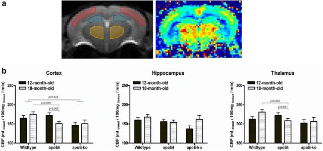
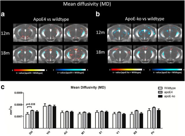
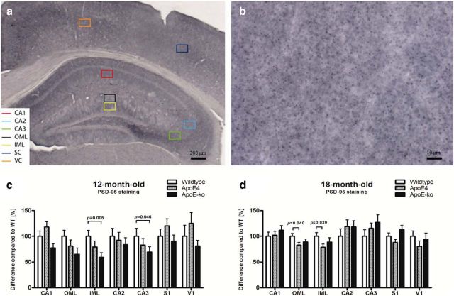
It is well established that the cholesterol-transporter apolipoprotein ε (APOE) genotype is associated with the risk of developing neurodegenerative diseases. Recently, brain functional connectivity (FC) in apoE-ε4 carriers has been investigated by means of resting-state fMRI, showing a marked differentiation in several functional networks at different ages compared with carriers of other apoE isoforms. The causes of such hampered FC are not understood. We hypothesize that vascular function and synaptic repair processes, which are both impaired in carriers of ε4, are the major contributors to the loss of FC during aging. To test this hypothesis, we integrated several different MRI techniques with immunohistochemistry and investigated FC changes in relation with perfusion, diffusion, and synaptic density in apoE4 and apoE-knock-out (KO) mice at 12 (adult) and 18 months of age. Compared with wild-type mice, we detected FC deficits in both adult and old apoE4 and apoE-KO mice. In apoE4 mice, these changes occurred concomitant with increased mean diffusivity in the hippocampus, whereas perfusion deficits appear only later in life, together with reduced postsynaptic density levels. Instead, in apoE-KO mice FC deficits were mirrored by strongly reduced brain perfusion since adulthood. In conclusion, we provide new evidence for a relation between apoE and brain connectivity, possibly mediated by vascular risk factors and by the efficiency of APOE as synaptic modulator in the brain. Our results show that multimodal MR neuroimaging is an excellent tool to assess brain function and to investigate early neuropathology and aging effects in translational research.

Keywords: apoE; apoE4 mice; cerebral blood flow; diffusion tensor imaging; functional connectivity; resting-state fMRI.


Figures

Figure 1.
Figure 1.
a, Anatomical locations of the cortical ROI, based on Ullmann et al., 2013. b, FC maps defined in the 12-month-old male wild-type mice by group ICA based on 30 components. The spatial color-coded z-maps of these components are overlaid on the template SE-EPI image. Components in same anatomical locations but in contralateral regions are shown together with two different color-codes, with a higher Z-score (yellow or light blue, thresholded for |Z| > 1.96) representing a higher correlation between the time course of that voxel and the mean time course for this component. Mean components comprise: M1–M2; M1–S1; M1–S1-V1; S1–V1; S2-AU; RS; piriform cortex (Pir); Cingulum (Cg); HC; CPu; Lateral septal nuclei (LS)-Th; Th-CPu, hypothalamus (HTc). c, By reducing the number of components to 15, many areas displayed an extended connectivity pattern across the same anatomical location, revealing a certain degree of interhemispheric connectivity. d, Other components having low amount of voxels at the edge of the brain, or displayed in only one slice, are considered artifacts.
Figure 2.
Figure 2.
Mean FC maps defined in three ROIs in wild-type, apoE4, and apoE-KO mice. Voxel-based FC maps of the different seeds-area positioned in the VH, somatosensory cortex (SS), and AU in the left hemisphere reveal functionally connected areas. The spatial color-coded FC maps are overlaid on the SE-EPI images of the same mouse. A higher p score (yellow) represents a higher correlation between the BOLD time course of the seed area and the other voxels in the brain. Voxels representing a significant reduction in FC (p < 0.05, minimum voxel cluster size 0.05 mm3) between apoE4, apoE-KO compared with wild-type are shown in blue. No voxels indicated an increased FC in these genotypes at both ages. a, At 12 months of age, a strong interhemispheric connectivity between right and left hippocampus is notable. In the cortical regions, the connectivity is covering SS, AU, and motor cortices, mainly unilaterally distributed but with extent also in contralateral areas, in the hippocampus and in few thalamic nuclei. VBA defined the location of significant FC reduction in the selected ROI in apoE4 and apoE-KO mice. b, At 18 months of age, the strength of correlation and the interhemispheric connectivity were visibly reduced but still present due to aging. Differences between genotypes were less visible and present mainly in the apoE-KO mice.
Figure 3.
Figure 3.
Resting-state FC based on total correlation analyses of 15 ROI in the mouse brain. a, Total correlation matrices of wild-type, apoE4 and apoE-KO mice at 12- and 18 months of age. Some characteristics of the mouse brain connectivity are consistent within different mouse models and age-effect; for example, it is visible a strong bilateral connectivity between DH and VH. AU, M1, S1, and RS also show a high degree of interconnectivity, as well as high FC between V1 (BPM; average ±SE). b, Statistical analyses of FC is shown as a matrix, to reveal significant differences between genotypes (bottom-right) and between aging (top-right) for each ROI–ROI connection. Because no genotype × age interactions were seen, data were analyzed as genotype effects, independent of age, and aging effects, independent of genotype. ApoE4 mice displayed a reduction of FC at both ages, compared with wild-type, between several regions; particularly, a reduced FC is seen between dorsal and ventral hippocampus and M1, AU, and SS. Intracortical FC reduction are also seen, affecting primarily the AU-M1-SS system. Aging effects are also shown, revealing a reduction of FC over time between 12- and 18-month-old mice. Most striking differences are found between M1 and DH, VH and RS. c, Statistical analyses revealed strong reduction of FC in apoE-KO mice compared with wild-type mice, at both ages. These deficits seem to occur primarily in the hippocampal-cortical connectivity, but also between M1, AU, S1, and RS. Reduced FC due to aging is seen between M1 and DH, and between AU cortices.
Figure 4.
Figure 4.
Resting-state FC based on partial correlation analyses of 17 ROIs in the mouse brain. a, Averaged partial correlation matrix of 12-month-old wild-type mice. Compared with total correlation analysis, the partial correlation highlights the direct connectivity between two regions, by regressing the temporal BOLD signal from all other ROI. These matrices were highly similar between different genotypes and ages (data not shown) and revealed unique features of mouse brain connectivity; in particular, a significant direct interhemispheric connectivity is seen within the DH, VH, AU, M1, and V1. Among the cortical regions, we could not detect a significant direct interhemispheric connectivity only in the S1 region. However, a strong intrahemispheric connectivity is seen between S1, M1, and AU and between V1 and RS. Resulting connectivity were thresholded at |Z| > 1.96, corresponding to uncorrected p < 0.025 for two-tailed test, based on the 12-month-old wild-type data. b, From the selected areas, reduced interhemispheric direct connectivity between auditory cortices was detected in apoE4 mice; significant reduced FC in the apoE-KO mice, independent of age, was seen between the S1 and the M1. Data represent mean ± SE.
Figure 5.
Figure 5.
a, CBF was measured in apoE4, apoE-KO, and wild-type mice at 12 and 18 months of age in three different ROIs: cortex (including somatosensory, auditory, and visual cortices), hippocampus, and thalamus. The area corresponding to the retrosplenial cortex was excluded, because it overlaps with the azygos pericallosal artery (azPA) of the anterior cerebral artery (ACA). To measure the CBF we used a FAIR ASL MRI technique. b, A reduced CBF was found in the cortex in 18-month-old apoE4 mice and in 12- and 18-month-old apoE-KO mice compared with wild-type. A similar trend was observed in the thalamus, although it was not significant for the apoE-KO mice. No significant differences were seen in the hippocampus.
Figure 6.
Figure 6.
MD comparison between wild-type, apoE4, and apoE-KO mice. VBA indicate significant differences in MD at 12 and 18 months of age. Three rostral to caudal averaged MD maps are overlaid with voxels that showed a significant difference (p < 0.05, minimum voxel cluster size 0.05 mm3). The voxel color indicates a negative or positive change in the apoE4 (a) or in the apoE-KO (b) mice compared with the wild-type mice of the same age. Few areas of significant differences are depicted in the explorative VBA; among them, an increase in MD in the DH of the apoE4 mice, independent of age, was also confirmed by ROI-based MANOVA (c). Increase in MD in the DH was also observed in the apoE-KO mice, although not significant (p > 0.05).
Figure 7.
Figure 7.
FA comparison between wild-type, apoE4 and apoE-KO mice. VBA indicate significant differences in FA at 12 and 18 months of age. Three rostral to caudal averaged FA maps are overlaid with voxels that showed a significant difference (p < 0.05, minimum voxel cluster size 0.05 mm3). The voxel color indicates a negative or positive change in the apoE4 (a) or in the apoE-KO (b) mice compared with the wild-type mice of the same age. GM regions did not show FA differences between groups (data not shown), although a reduced FA is visible in the molecular layer of the hippocampus in apoE4 mice, as depicted in the explorative VBA (a). ROI analyses of the CC in different bregma did not show statistical significant differences between FA due to genotype or aging (c).
Figure 8.
Figure 8.
PSD-95 staining performed on brain sections of apoE-ε4, apoE-KO, and wild-type mice. a, b, Representation of PSD-95-immunoreactive staining in hippocampal and cortical areas at two different magnifications (5× and 100×, respectively). c, In 12-month-old animals we detected a significant decrease in PSD-95 levels in apoE-KO mice compared with wild-type in the IML and CA3 of hippocampus. d, In 18-month-old animals we show reduced PSD-95 levels in the apoE-ε4 animals in the OML and in the IML, whereas no significant differences were seen between wild-type and apoE-KO mice. Values represent the mean and SEM.

Similar articles

Cited by

References

    1. Biswal B, Yetkin FZ, Haughton VM, Hyde JS. Functional connectivity in the motor cortex of resting human brain using echo-planar MRI. Magn Reson Med. 1995;34:537–541. doi: 10.1002/mrm.1910340409. - DOI - PubMed
    1. Blain JF, Sullivan PM, Poirier J. A deficit in astroglial organization causes the impaired reactive sprouting in human apolipoprotein E4 targeted replacement mice. Neurobiol Dis. 2006;21:505–514. doi: 10.1016/j.nbd.2005.08.010. - DOI - PubMed
    1. Bondi MW, Houston WS, Eyler LT, Brown GG. fMRI evidence of compensatory mechanisms in older adults at genetic risk for Alzheimer disease. Neurology. 2005;64:501–508. doi: 10.1212/01.WNL.0000150885.00929.7E. - DOI - PMC - PubMed
    1. Bookheimer SY, Strojwas MH, Cohen MS, Saunders AM, Pericak-Vance MA, Mazziotta JC, Small GW. Patterns of brain activation in people at risk for Alzheimer's disease. N Engl J Med. 2000;343:450–456. doi: 10.1056/NEJM200008173430701. - DOI - PMC - PubMed
    1. Brown JA, Terashima KH, Burggren AC, Ercoli LM, Miller KJ, Small GW, Bookheimer SY. Brain network local interconnectivity loss in aging APOE-4 allele carriers. Proc Natl Acad Sci U S A. 2011;108:20760–20765. doi: 10.1073/pnas.1109038108. - DOI - PMC - PubMed

Publication types