. 2014 Dec 5;289(49):34089-103.
doi: 10.1074/jbc.M114.607168. Epub 2014 Oct 15.

A role for versican in the development of leiomyosarcoma

Affiliations

A role for versican in the development of leiomyosarcoma

Paul A Keire et al. J Biol Chem. .

Abstract

Leiomyosarcoma (LMS) is a mesenchymal cancer that occurs throughout the body. Although LMS is easily recognized histopathologically, the cause of the disease remains unknown. Versican, an extracellular matrix proteoglycan, increases in LMS. Microarray analyses of 80 LMSs and 24 leiomyomas showed a significant elevated expression of versican in human LMS versus benign leiomyomas. To explore the importance of versican in this smooth muscle cell tumor, we used versican-directed siRNA to knock down versican expression in a LMS human cell line, SK-LMS-1. Decreased versican expression was accompanied by slower rates of LMS cell proliferation and migration, increased adhesion, and decreased accumulation of the extracellular matrix macromolecule hyaluronan. Addition of purified versican to cells expressing versican siRNA restored cell proliferation to the level of LMS controls, increased the pericellular coat and the retention of hyaluronan, and decreased cell adhesion in a dose-dependent manner. The presence of versican was not only synergistic with hyaluronan in increasing cell proliferation, but the depletion of versican decreased hyaluronan synthase expression and decreased the retention of hyaluronan. When LMS cells stably expressing versican siRNA were injected into nude mice, the resulting tumors displayed significantly less versican and hyaluronan staining, had lower volumes, and had reduced levels of mitosis as compared with controls. Collectively, these results suggest a role for using versican as a point of control in the management and treatment of LMS.

Keywords: Cell Migration; Cell Proliferation; Extracellular Matrix; Hyaluronan; Leiomyosarcoma; Small Interfering RNA (siRNA); Tumor Growth; Versican.


Figures

FIGURE 1.
FIGURE 1.
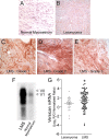
Versican is highly expressed in clinical samples of leiomyosarcoma tissue. Histological assessment of normal myometrium shows low to undetectable levels of versican expression (A) and an increase in versican levels in leiomyomas (B) when compared with normal myometrium. This increase, however, is intermediate and less than that found in grade 1, 2, and 3 leiomyosarcoma tumors. The three grades of tumor (LMS grades 1–3; C–E) express comparably more versican. Grade 1 and grade 2 LMS tumors (C and D) displayed the greatest versican expression in terms of both intensity and distribution. The increased cellularity in lower grade tumors appeared to correspond to areas of high versican expression. Original magnification, 400×. F, Northern blot analyses of LMS tissue show a high level of versican transcript versus adjacent normal myometrial controls. The blot represents loading based on equal amounts of 28 S RNA. Arrowheads (◁) indicate versican isoform bands V0 and V1 on the basis of size, top to bottom (79). G, versican mRNA is up-regulated in LMS versus benign leiomyoma. Scatter plots are shown for versican mRNA expression levels in clinical samples of 80 LMSs and 24 benign leiomyomas as analyzed by gene expression microarrays. On average, LMS samples showed an approximate 2-fold increase in versican transcript levels when compared with leiomyoma samples, which represented a statistically significant up-regulation, R/G, red-to-green ratio (mean ± S.E. (error bars); t test, *, p = 0.0365).
FIGURE 2.
FIGURE 2.
Versican siRNA efficiently inhibits versican expression. By constitutive expression of versican siRNA in LMS cells, as much as an 85% reduction in versican mRNA (A) and a 95% reduction in versican protein were achieved (B). * represents statistical significance (p < 0.05) between LMS/siRNA Vc clones (1, 2, and 3) compared with LMS control cells (error bars represent ±S.D.). Densitometry of the Western blot (B) depicts the relative versican protein levels for the V1 isoform. C and D, LMS/EV or LMS/siRNA Vc cells were cultured for 24 h on 8-well chamber glass slides. Cells were fixed and immunocytochemically stained for versican (red). Cell nuclei were counterstained with Hoechst 33342 (blue). Immunocytochemical analysis confirmed that versican levels were significantly reduced in the LMS/siRNA Vc cells.
FIGURE 3.
FIGURE 3.
LMS/siRNA Vc cells are more polygonal, have a less migratory phenotype, and are more adhesive to cell culture plastic compared with LMS cells. A, LMS/EV (shown), LMS/WT, and LMS/siRNA Scr control cells growing from clonal islands assume spindle shapes typical of rapidly proliferating smooth muscle cells. B, in contrast, LMS cells transduced to express versican siRNA are flatter, more polygonal, and cluster into aggregates. Scale bars, 30 μm. C, trypsinization time for complete removal of the cells was ∼10 times longer for the versican siRNA cells (gray bar) than the LMS/EV controls (black bar) (n = 6) (***, p < 0.0001). D, addition of versican to the LMS/siRNA Vc cells reversed the adhesive phenotype in a dose-dependent manner. Error bars represent ±S.E.
FIGURE 4.
FIGURE 4.
DNA replication, cell proliferation, and migration are inhibited by versican knockdown; however, cell proliferation is reversible by versican add-back. [3H]Thymidine levels (A) and cell proliferation (B) indicate that the LMS/WT (●) and LMS/siRNA Scr (▾) control cells divide and proliferate at a significantly higher rate than the two different versican siRNA LMS cells (□ and ○). C, the reduction in the proliferative rate induced by versican siRNA can be reversed by the addition of purified versican to the culture medium. Exogenous versican exerts its effect in a dose-dependent manner (*, p < 0.05; analyzed at 40 h). D, in a wound migration assay, the migration of LMS cells (▴) was significantly greater (*, p < 0.05) at 12 and 24 h than that of LMS cells transduced with versican siRNA (○) (n = 4). Error bars represent ±S.E.
FIGURE 5.
FIGURE 5.
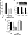
Reduction in hyaluronan and HAS levels with a reduction in versican synthesis. A, a decrease in hyaluronan production levels followed versican levels knocked down by siRNA. LMS/siRNA Vc clones (gray bars) along with normal uterine smooth muscle cell (NUtSMC) controls (open bar) produce significantly less total hyaluronan than the LMS/WT tumor cells (black bar) and on an equal cell basis (*, p < 0.05). B, RT-PCR for HAS2, HAS3, and versican for LMS/WT control tumor cells and LMS/siRNA Vc cells (upper panel) in which versican mRNA had been constitutively reduced using versican-directed siRNA. Densitometry of RT-PCR averaged control (n = 4) (black bars) or siRNA clones (n = 3) (gray bars) showed a significant reduction in the levels of HAS2, HAS3, and versican in the cells expressing versican siRNA (*, p < 0.05). All lanes were normalized to GAPDH. C, large molecular weight hyaluronan by itself does not restore the proliferative profile of LMS/siRNA Vc cells to LMS/WT levels but does with the addition of purified versican. Although there is a significant increase in cell proliferation with the addition of hyaluronan (30 μg/ml; *, p < 0.015), the increase due to the addition of versican at nanogram levels is significantly greater (***, p < 0.0001), and near complete restoration (96.6%) of the cell native proliferative rate is achieved at 100 μg/ml versican. The difference between versican alone and versican + large hyaluronan is significant (**, p < 0.004), suggesting an additive or synergistic effect between hyaluronan and versican on cell proliferation. Error bars represent ±S.D.
FIGURE 6.
FIGURE 6.
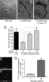
Versican increases pericellular coat thickness and limits hyaluronan degradation. A, LMS/WT (shown), LMS/EV, and LMS/siRNA Scr control cells have prominent pericellular coats. Arrowheads (▷) and dotted white lines mark the pericellular boundaries. B, LMS/siRNA Vc cells display a diminished cell coat, which increases in thickness with the addition of exogenous versican (C). Individual cells were examined with a particle exclusion assay 24 h after the addition of versican. Measurements were perpendicular to the cell membrane at the point of maximum coat thickness. Scale bars, 50 μm. The cell coat thickness of the LMS cells is significantly greater than the LMS/siRNA Vc cells (*, p < 0.05) and increased significantly (*, p < 0.05) with the addition of exogenous versican to the cell medium. D, bar graph representing the average ± S.E. of n = 172 cells counted for control LMS cells, n = 293 LMS/siRNA Vc cells with 0 μg/ml versican, n = 104 cells with 3 μg/ml versican, and n = 67 cells with 10 μg/ml versican. E–G, degradation of internalized FITC-hyaluronan is inhibited in the presence of versican. Media for versican-depleted LMS/siRNA Vc cells was supplemented with FITC-labeled hyaluronan for 72 h, and then the cells were trypsinized and replated. After 24 h, the amount of FITC-labeled hyaluronan was measured in the absence (E) or presence (F) of 10 μg/ml purified versican. Scale bars, 100 μm. Cells receiving versican retained a significantly greater amount of hyaluronan (*, p < 0.001) (G). Error bars represent ±S.D.
FIGURE 7.
FIGURE 7.
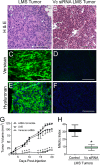
Versican and hyaluronan are highly expressed in LMS tumors in comparison with versican siRNA tumors. LMS tumors also grow continuously with a high mitotic index, whereas versican siRNA tumors grow only slowly and at one-third the mitotic index. Images (A–F) represent subcutaneous tumors excised after 21 days in nude mice; all image pairs were taken at equal exposure. H&E staining of tumor sections reflects a highly hyperplastic cellular response in the LMS tumors (A) not seen in the versican siRNA tumors (B). Multinucleation, cytologic pleomorphisms (arrowheads), and tissue necrosis were observed in LMS tumors, whereas less cellular atypia and no evidence of necrosis were observed in the versican siRNA tumors. Scale bars, 100 μm. Original magnification, 400×. The constitutively expressing versican siRNA LMS cells (panels on the right) only faintly express versican (D) and hyaluronan (F) compared with the tumors of the empty vector control panels on the left (C, versican; E, hyaluronan). Over the 3-week study period, the LMS tumors grew continuously (G) with a high mitotic index (H), whereas versican siRNA tumors grew slowly and at one-third the mitotic index. G, tumor volume over time in nude mice injected subcutaneously in LMS/EV (▴; n = 5; ±S.E.) and LMS/siRNA Scr (▾; n = 5; ±S.E.) control cell tumors versus LMS/siRNA Vc cell tumors (○; n = 11; ±S.E.). *, p < 0.001. Error bars, ±S.E. H, mitotic index of LMS control cell tumors versus LMS/siRNA Vc cell tumors. The box graph depicts median ± S.D., and error bars show the minimum and maximum range of mitotic figures per 10 400× fields (n = 15). *, p < 0.01.
FIGURE 8.
FIGURE 8.
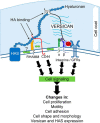
Schematic diagram that details the described interplay of versican and hyaluronan in LMS tumorigenesis. RHAMM, receptor for hyaluronan-mediated motility; GFRs, growth factor receptors; HA, hyaluronan.

Similar articles

Cited by

References

    1. Major F. J., Blessing J. A., Silverberg S. G., Morrow C. P., Creasman W. T., Currie J. L., Yordan E., Brady M. F. (1993) Prognostic factors in early-stage uterine sarcoma. A Gynecologic Oncology Group study. Cancer 71, 1702–1709 - PubMed
    1. Moinfar F., Azodi M., Tavassoli F. A. (2007) Uterine sarcomas. Pathology 39, 55–71 - PubMed
    1. Zako M., Shinomura T., Ujita M., Ito K., Kimata K. (1995) Expression of PG-M (V3), an alternatively spliced form of PG-M without a chondroitin sulfate attachment region in mouse and human tissues. J. Biol. Chem. 270, 3914–3918 - PubMed
    1. Wight T. N., Kinsella M. G., Evanko S. P., Potter-Perigo S., Merrilees M. J. (2014) Versican and the regulation of cell phenotype in disease. Biochim. Biophys. Acta 1840, 2441–2451 - PMC - PubMed
    1. Lemire J. M., Braun K. R., Maurel P., Kaplan E. D., Schwartz S. M., Wight T. N. (1999) Versican/PG-M isoforms in vascular smooth muscle cells. Arterioscler. Thromb. Vasc. Biol. 19, 1630–1639 - PubMed

Publication types

MeSH terms

LinkOut - more resources