. 2014 Nov 21;289(47):33054-65.
doi: 10.1074/jbc.M114.582304. Epub 2014 Oct 15.

KRIT1 protein depletion modifies endothelial cell behavior via increased vascular endothelial growth factor (VEGF) signaling

Affiliations

KRIT1 protein depletion modifies endothelial cell behavior via increased vascular endothelial growth factor (VEGF) signaling

Peter V DiStefano et al. J Biol Chem. .

Abstract

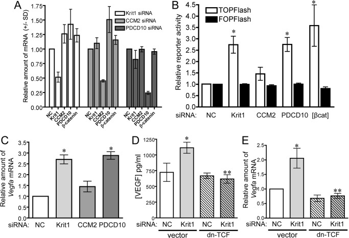
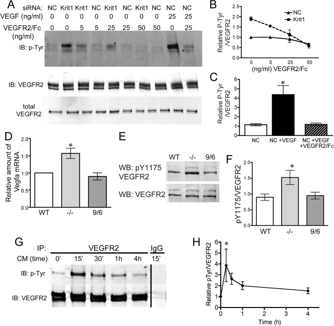
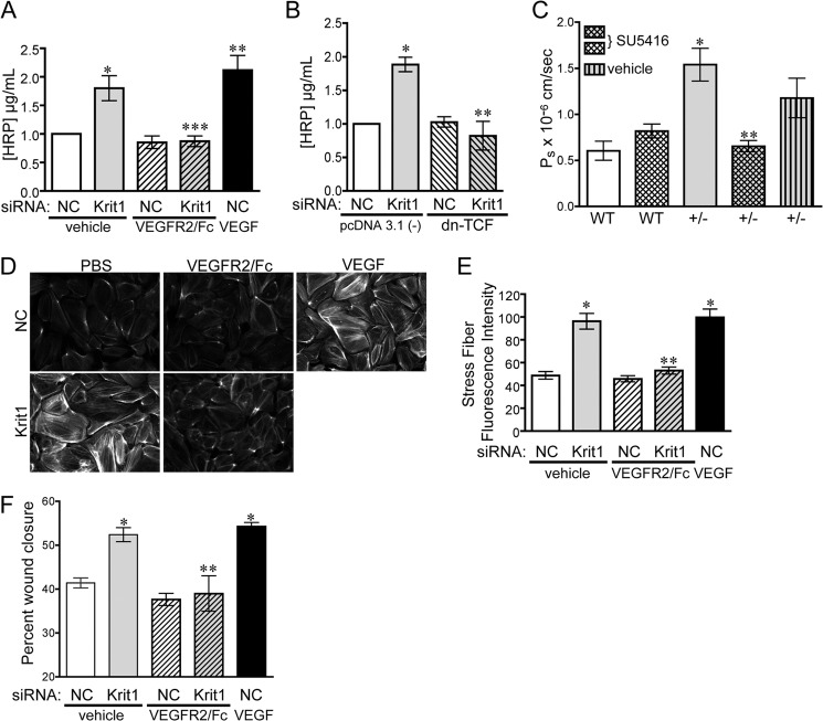
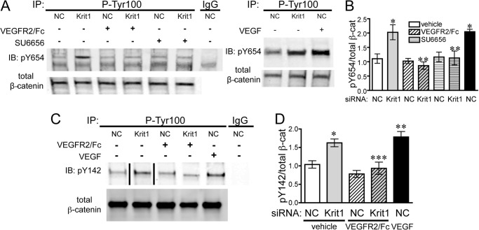
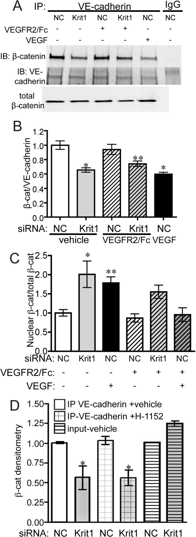
Disruption of endothelial cell-cell contact is a key event in many cardiovascular diseases and a characteristic of pathologically activated vascular endothelium. The CCM (cerebral cavernous malformation) family of proteins (KRIT1 (Krev-interaction trapped 1), PDCD10, and CCM2) are critical regulators of endothelial cell-cell contact and vascular homeostasis. Here we show novel regulation of vascular endothelial growth factor (VEGF) signaling in KRIT1-depleted endothelial cells. Loss of KRIT1 and PDCD10, but not CCM2, increases nuclear β-catenin signaling and up-regulates VEGF-A protein expression. In KRIT1-depleted cells, increased VEGF-A levels led to increased VEGF receptor 2 (VEGFR2) activation and subsequent alteration of cytoskeletal organization, migration, and barrier function and to in vivo endothelial permeability in KRIT1-deficient animals. VEGFR2 activation also increases β-catenin phosphorylation but is only partially responsible for KRIT1 depletion-dependent disruption of cell-cell contacts. Thus, VEGF signaling contributes to modifying endothelial function in KRIT1-deficient cells and microvessel permeability in Krit1(+/-) mice; however, VEGF signaling is likely not the only contributor to disrupted endothelial cell-cell contacts in the absence of KRIT1.

Keywords: Adherens Junction; CCM; Krit1; Permeability; Phosphotyrosine Signaling; Vascular Endothelial Growth Factor (VEGF); β-Catenin (B-Catenin).


Figures

FIGURE 1.
FIGURE 1.
Loss of KRIT1 and PDCD10, but not CCM2, stimulates nuclear β-catenin-dependent expression of Vegfa. A, knockdown efficiency of anti-KRIT1, CCM2, and PDCD10 siRNA on Krit1, CCM2, PDCD10, and Ctnnb1-catenin) mRNA expression. B, β-catenin-dependent reporter activity in HPAEC transfected with NC, anti-KRIT1, -CCM2, and -PDCD10 siRNA. TOPFlash activity is shown relative to NC siRNA-transfected cells (±S.E.) and compared with cells overexpressing β-catenin [βcat]. n = 4, p = 0.0011 by ANOVA; *, p < 0.05 by post-hoc test versus NC-transfected cells. C, expression of Vegfa mRNA in siRNA-transfected HPAEC. Data shown are the average expression from three experiments, ± S.E., p = 0.0002 by ANOVA; *, p < 0.001 by post-hoc test versus NC-transfected cells. D, concentration of VEGF-A secreted from NC and KRIT1 siRNA-transfected cells. n = 9, p = 0.003 by ANOVA; *, p < 0.05 by post-hoc test versus NC-transfected cells; **, p < 0.01 versus KRIT1 siRNA-transfected cells. E, expression of Vegfa mRNA in siRNA-transfected cells co-expressing dn-TCF ±S.E; n = 3, p = 0.003 by ANOVA; *, p < 0.05 by post-hoc test versus NC-transfected cells; **, p < 0.01 versus KRIT1 siRNA-transfected cells.
FIGURE 2.
FIGURE 2.
Expression of VEGF by KRIT1-depleted cells activates VEGFR2. A, tyrosine phosphorylation of VEGFR2 in NC- and KRIT1-siRNA-transfected cells treated with 5, 25, or 50 ng/ml VEGFR2/Fc ± rhVEGF (25 ng/ml). IB, immunoblot. Blots are representative, n = 4. B, densitometric quantification of VEGFR2 phosphorylation from four independent experiments treated as in A. Tyr(P) (P-Tyr) signal was normalized to VEGFR2 immunoprecipitation, shown relative to untreated NC-transfected cells, and plotted versus VEGFR2/Fc ± S.E. n = 4 independent experiments. C, densitometric quantification of VEGFR2 tyrosine phosphorylation after VEGF treatment from four independent experiments treated as in A. Tyr(P) signal was normalized to VEGFR2 immunoprecipitation and shown relative to untreated NC-transfected cells ± S.E. p < 0.0001 by ANOVA; *, p < 0.05 by post-hoc testing versus untreated NC-transfected cells. D, expression of Vegfa mRNA in WT, Krit1 null (−/−), and Krit1 reconstituted (9/6) MEF; ±S.E; n = 3; p = 0.008 by ANOVA. *, p < 0.05 by post-hoc test versus WT. E, tyrosine 1175 phosphorylation (pY1175) in WT, −/−, and 9/6 MEF. WB, Western blot. n = 7. F, densitometric quantification of VEGFR2 Tyr(P)-1175 for all experiments treated as in E. Data shown are Tyr(P)-1175 normalized to total VEGFR2, ± S.E; n = 7; p = 0.02 by ANOVA; *, p < 0.05 by post-hoc testing versus WT. G, tyrosine phosphorylation of VEGFR2 in NC siRNA-transfected cells treated with conditioned media from KRIT1-siRNA-transfected cells. CM, conditioned media. Blots are representative, n = 3. IP, immunoprecipitation. H, densitometric quantification of VEGFR2 tyrosine phosphorylation from 3 independent experiments treated as in G. Tyr(P) signal was normalized to VEGFR2 immunoprecipitation and plotted versus time, ±S.E; p = 0.0023 by ANOVA; *, p < 0.01 by post-hoc testing versus untreated.
FIGURE 3.
FIGURE 3.
Loss of KRIT1 leads to VEGF-dependent barrier disruption and migration. A, HRP leak through BAEC monolayers transfected with NC- or KRIT1-siRNA ± 25 ng/ml VEGFR2/Fc or recombinant human VEGF (50 ng/ml). Data shown are the mean HRP concentration, ± S.E. n = 3, p = 0.0005 by ANOVA; *, p < 0.05 by post-hoc testing versus NC-transfected cells; **, p < 0.01 versus NC-transfected cells; ***, p < 0.01 versus KRIT1. B, HRP leak through BAEC monolayers transfected with NC- or KRIT1-siRNA, ± co-expression of dn-TCF. Data shown are the mean HRP concentration ± S.E; n = 3; p = 0.001 by ANOVA; *, p < 0.01 by post-hoc test versus NC; **, p < 0.01 versus KRIT1 siRNA-transfected cells. C, cremaster microvessel permeability in WT or Krit1+/− (+/-) mice treated ± 3 mg/kg SU5416 or with vehicle alone. Data shown are the mean Ps ± S.E. n ≥ 17 vessel sites; p < 0.0001 by non-parametric ANOVA; *, p < 0.001 by post-hoc testing versus WT; **, p < 0.001 versus +/−. D, epifluorescence images of rhodamine-phalloidin-stained NC and KRIT1-siRNA-transfected HPAE-treated ± 25 ng/ml VEGFR2/Fc or 50 ng/ml rhVEGF. Images are representative of three separate experiments; scale bar = 50 μm. E, fluorescent intensity quantification of D. n ≥ 40 cells from 10 fields of view ±S.E. p < 0.0001 by ANOVA; *, p < 0.001 by post-hoc test versus NC-transfected cells; **, p < 0.001 versus KRIT1 siRNA-transfected cells. F, percent wound closure of NC- and KRIT1-siRNA-transfected cells treated ± 25 ng/ml VEGFR2/Fc or 25 ng/ml rhVEGF. Data shown are the mean ± S.E. n = 8; p < 0.0001 by ANOVA; *, p < 0.001 by post-hoc test versus NC-transfected cells; **, p < 0.001 versus KRIT1-siRNA-transfected cells.
FIGURE 4.
FIGURE 4.
KRIT1 depletion leads to VEGF-dependent tyrosine phosphorylation of β-catenin. A, left panel, tyrosine 654 phosphorylation (pY654) of β-catenin in NC- and KRIT1-siRNA-transfected BAEC-treated ± 25 ng/ml VEGFR2/Fc or ± 1 μm SU6656. IgG, mouse IgG; IP, immunoprecipitation; IB, immunoblot. Right panel, comparison of KRIT1 siRNA-induced and VEGF-induced (50 ng/ml) Tyr-654 phosphorylation. Blots are representative, n = 4. B, densitometric quantification of β-catenin Tyr-654 phosphorylation from four independent experiments treated as in A. Tyr(P)-654 signal was normalized to β-catenin immunoprecipitation and displayed relative to NC-transfected cells, ±S.E. p = 0.002 by ANOVA; *, p < 0.01 by post-hoc testing versus NC-transfected cells; **, p < 0.05 versus KRIT1 siRNA-transfected cells. C, tyrosine 142 phosphorylation (pY142) of β-catenin in NC- and KRIT1-siRNA-transfected BAEC-treated ± 25 ng/ml VEGFR2/Fc or 50 ng/ml rhVEGF. The line indicates removal of intervening lanes. Blots are representative, n = 3. D, densitometric quantification of β-catenin Tyr-142 phosphorylation from three independent experiments treated as in C. The Tyr(P)-142 signal was normalized to β-catenin immunoprecipitation and displayed relative to NC-transfected cells ±S.E. p = 0.0003 by ANOVA; *, p < 0.05 by post-hoc testing versus NC-transfected cells; **, < 0.01 versus NC-transfected cells; ***, p < 0.05 versus KRIT1 siRNA-transfected cells.
FIGURE 5.
FIGURE 5.
KRIT1 depletion-dependent dissociation of β-catenin and VE-cadherin is not reversed by inhibition of VEGFR2. A, co-immunoprecipitation of VE-cadherin and β-catenin in NC- and KRIT1-siRNA-transfected HPAEC-treated ± 25 ng/ml VEGFR2/Fc or rhVEGF (50 ng/ml). IgG, rabbit IgG; IP, immunoprecipitation; IB, immunoblot. Blots are representative, n = 5. B, densitometric quantification of β-catenin co-immunoprecipitation from five independent experiments treated as in A. Data shown are β-catenin-normalized to VE-cadherin immunoprecipitation ±S.E. p = 0.0004 by ANOVA; *, p < 0.001; **, p < 0.05 by post-hoc testing versus untreated NC-transfected cells. C, densitometric quantification of nuclear β-catenin in NC- and KRIT1-siRNA-transfected HPAEC-treated ± 50 ng/ml VEGFR2/Fc or rhVEGF (50 ng/ml). Nuclear fractions were isolated as described under “Experimental Procedures.” Data shown are nuclear β-catenin-normalized to total β-catenin for each condition from 5 independent experiments. p = 0.0013 by ANOVA; *, p < 0.01; **, p < 0.05 by Dunnett's multiple comparison test versus untreated NC-transfected cells. D, densitometric quantification of β-catenin co-immunoprecipitation with VE-cadherin in NC and KRIT1 siRNA-transfected HPAE treated with 100 nm H-1152. Data are combined from five independent experiments. Data shown are β-catenin-normalized to VE-cadherin immunoprecipitation ±S.E. p = 0.0082 by ANOVA; *, p < 0.05 by post-hoc testing versus vehicle-treated NC-transfected cells.

Similar articles

Cited by

References

    1. Wallez Y., Huber P. (2008) Endothelial adherens and tight junctions in vascular homeostasis, inflammation, and angiogenesis. Biochim. Biophys. Acta 1778, 794–809 - PubMed
    1. Orr A. W., Stockton R., Simmers M. B., Sanders J. M., Sarembock I. J., Blackman B. R., Schwartz M. A. (2007) Matrix-specific p21-activated kinase activation regulates vascular permeability in atherogenesis. J. Cell Biol. 176, 719–727 - PMC - PubMed
    1. Mammoto A., Mammoto T., Kanapathipillai M., Wing Yung C., Jiang E., Jiang A., Lofgren K., Gee E. P., Ingber D. E. (2013) Control of lung vascular permeability and endotoxin-induced pulmonary oedema by changes in extracellular matrix mechanics. Nat. Commun. 4, 1759. - PubMed
    1. Lee Y. C. (2005) The involvement of VEGF in endothelial permeability: a target for anti-inflammatory therapy. Curr. Opin. Investig. Drugs 6, 1124–1130 - PubMed
    1. Middleton J., Americh L., Gayon R., Julien D., Aguilar L., Amalric F., Girard J. P. (2004) Endothelial cell phenotypes in the rheumatoid synovium: activated, angiogenic, apoptotic and leaky. Arthritis Res. Ther. 6, 60–72 - PMC - PubMed

Publication types

MeSH terms