

Osteomyelitis of the patella: ensure a high index of suspicion and beware the negative aspirate
- PMID: 25320263
- PMCID: PMC4202083
- DOI: 10.1136/bcr-2014-206630
Osteomyelitis of the patella: ensure a high index of suspicion and beware the negative aspirate
Abstract
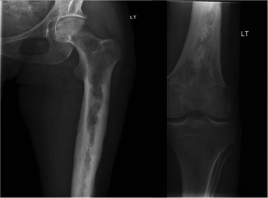
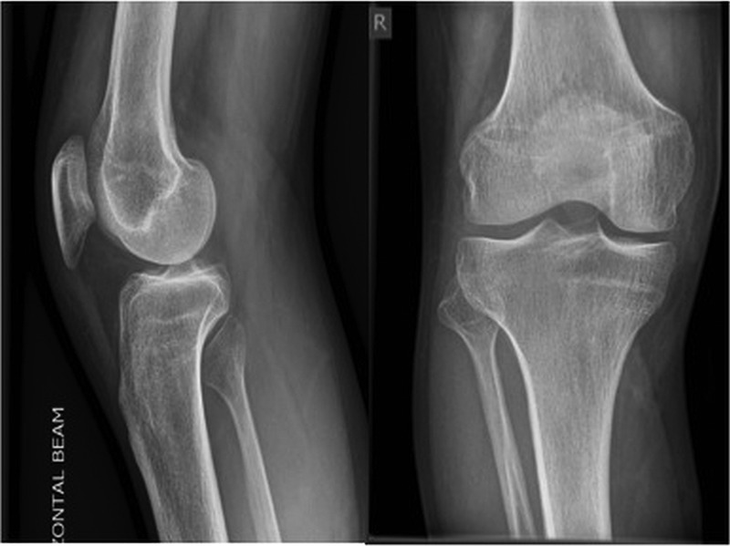
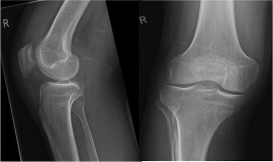
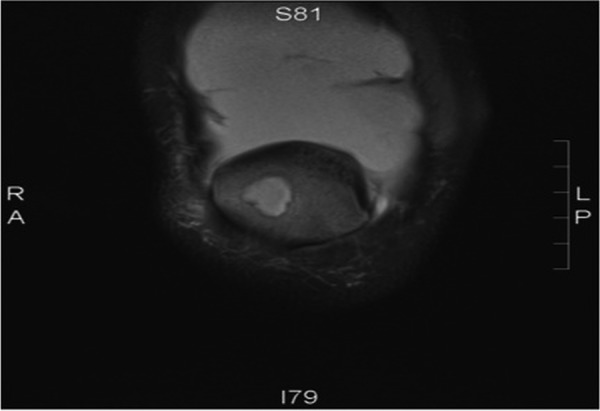
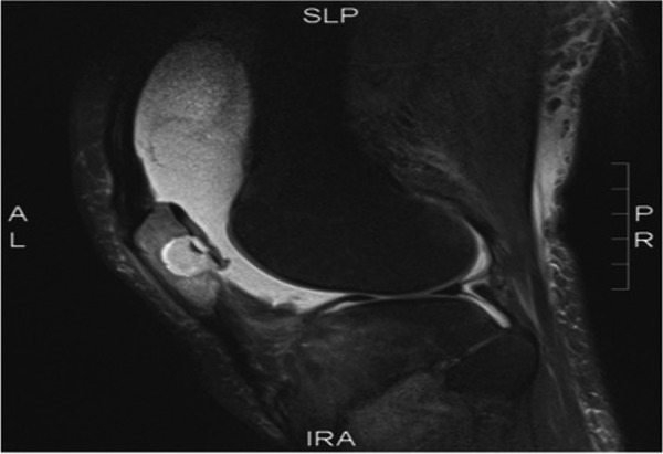
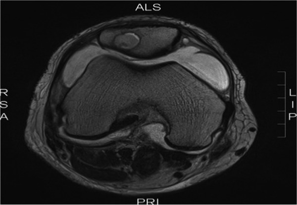
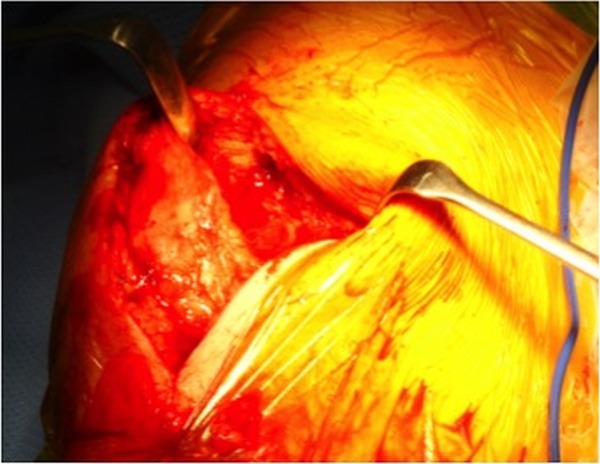
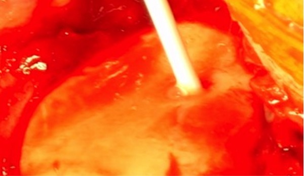
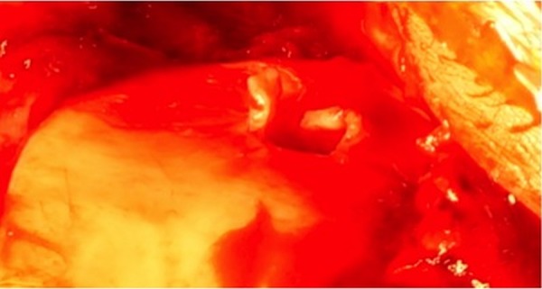
We report the case of a 33-year-old man who presented several times to healthcare professionals over a 6-week period with a painful swollen right knee. He had a history of chronic osteomyelitis of the left femur and had recently stopped taking suppressive antibiotics. A joint aspirate did not demonstrate any organisms. On subsequent review by the orthopaedic team MRI was performed which revealed an isolated area of osteomyelitis and an abscess in his right patella. He underwent arthrotomy, debridement and irrigation of the joint alongside antibiotic treatment. We highlight this case, as isolated osteomyelitis of the patella is a rare condition, especially in adults. In addition, the presenting features of osteomyelitis of the patella are varied and joint fluid aspirates often do not reveal an organism. This case therefore aims to raise an awareness of this condition and thereby ensure a high index of suspicion when symptoms or signs are present and inform clinicians of the investigative steps in order to avoid a delay in diagnosis as seen in this case.
2014 BMJ Publishing Group Ltd.
Figures

References
-
- Smith IM, Austin OMB, Batchelor AG. The treatment of chronic osteomyelitis: a 10 year audit. J Plast Reconstr Aesthet Surg 2006;59:11–15 - PubMed
-
- Pineda C, Vargas A, Rodriguez AV. Imaging of osteomyelitis: current concepts. Infect Dis Clin North Am 2006;20:789–825 - PubMed
-
- Sia IG, Berbari EF. Infection and musculoskeletal conditions: osteomyelitis. Best Pract Res Clin Rheumatol 2006;20:1065–81 - PubMed
-
- Lew DP, Waldvogel FA. Osteomyelitis. Lancet 2004;364:369–79 - PubMed
Publication types
MeSH terms
Substances
LinkOut - more resources
Full Text Sources
Other Literature Sources