. 2015 Jan;89(1):144-54.
doi: 10.1128/JVI.02308-14. Epub 2014 Oct 15.

Rhabdovirus-based vaccine platforms against henipaviruses

Affiliations

Rhabdovirus-based vaccine platforms against henipaviruses

Drishya Kurup et al. J Virol. 2015 Jan.

Abstract

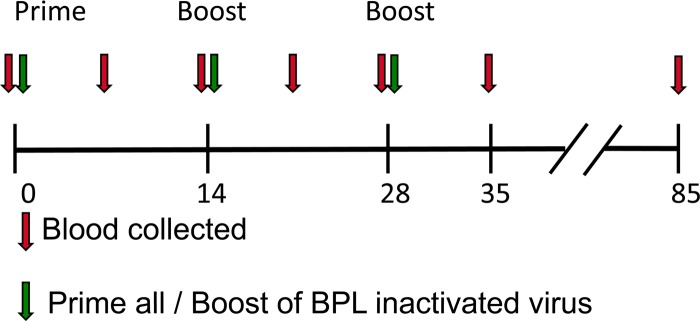
The emerging zoonotic pathogens Hendra virus (HeV) and Nipah virus (NiV) are in the genus Henipavirus in the family Paramyxoviridae. HeV and NiV infections can be highly fatal to humans and livestock. The goal of this study was to develop candidate vaccines against henipaviruses utilizing two well-established rhabdoviral vaccine vector platforms, recombinant rabies virus (RABV) and recombinant vesicular stomatitis virus (VSV), expressing either the codon-optimized or the wild-type (wt) HeV glycoprotein (G) gene. The RABV vector expressing the codon-optimized HeV G showed a 2- to 3-fold increase in incorporation compared to the RABV vector expressing wt HeV G. There was no significant difference in HeV G incorporation in the VSV vectors expressing either wt or codon-optimized HeV G. Mice inoculated intranasally with any of these live recombinant viruses showed no signs of disease, including weight loss, indicating that HeV G expression and incorporation did not increase the neurotropism of the vaccine vectors. To test the immunogenicity of the vaccine candidates, we immunized mice intramuscularly with either one dose of the live vaccines or 3 doses of 10 μg chemically inactivated viral particles. Increased codon-optimized HeV G incorporation into RABV virions resulted in higher antibody titers against HeV G compared to inactivated RABV virions expressing wt HeV G. The live VSV vectors induced more HeV G-specific antibodies as well as higher levels of HeV neutralizing antibodies than the RABV vectors. In the case of killed particles, HeV neutralizing serum titers were very similar between the two platforms. These results indicated that killed RABV with codon-optimized HeV G should be the vector of choice as a dual vaccine in areas where rabies is endemic.

Importance: Scientists have been tracking two new viruses carried by the Pteropid fruit bats: Hendra virus (HeV) and Nipah virus (NiV). Both viruses can be fatal to humans and also pose a serious risk to domestic animals. A recent escalation in the frequency of outbreaks has increased the need for a vaccine that prevents HeV and NiV infections. In this study, we performed an extensive comparison of live and killed particles of two recombinant rhabdoviral vectors, rabies virus and vesicular stomatitis virus (VSV), expressing wild-type or codon-optimized HeV glycoprotein, with the goal of developing a candidate vaccine against HeV. Based on our data from the presented mouse immunogenicity studies, we conclude that a killed RABV vaccine would be highly effective against HeV infections and would make an excellent vaccine candidate in areas where both RABV and henipaviruses pose a threat to human health.


Figures

FIG 1
FIG 1
RABV- and VSV-based vaccine vectors. Shown is a schematic representation of the parental vectors (RABV and VSV) and the vaccine constructs expressing wild-type HeV G [RABV-HeVG (wt) and VSV-HeVG (wt)] or codon-optimized HeV G (RABV-coHeVG and VSV-coHeVG).
FIG 2
FIG 2
FACS analysis of expression of viral glycoproteins. Vero cells were infected with the recombinant RABVs or VSVs as indicated. Forty-eight hours (RABV) or 6.5 h (VSV) after infection, cells were stained with antibodies directed against RABV G, VSV G, or HeV G, as indicated, and analyzed by FACS. The numbers indicate the mean fluorescence intensity (MFI) for each staining.
FIG 3
FIG 3
Analysis of purified virions of vaccine vectors. Purified virions (Vero cell derived) from RABV-coHeVG, RABV-HeVG, RABV, VSV-coHeVG, VSV-HeVG, or VSV-GFP were separated by SDS-PAGE and stained with SYPRO Ruby for total protein analysis (A) or transferred to a nitrocellulose membrane, and HeV G was detected by Western blotting (B). Lane M, molecular mass markers.
FIG 4
FIG 4
Pathogenicity study in Swiss Webster mice. Shown are weight curves of infected mice (Ms 1 to Ms 5) after i.n. inoculation with 105 PFU VSV or 105 FFU RABV live virus as indicated.
FIG 5
FIG 5
Experimental timeline of immunization study. BALB/c mice were immunized with either one dose of 105 PFU of live virus (day 0) or 3 doses of 10 μg inactivated virus (days 0, 14, and 28). Serum was collected every 7 days until day 35 and analyzed by ELISA. Final serum samples were collected on day 85 and used for determination of antibody neutralization titers.
FIG 6
FIG 6
Analysis of anti-HeV G humoral immune response of immunized mice. BALB/c mice were immunized as indicated in the legend to Fig. 5, and blood was collected on days 0, 7, 14, 21, 28, and 35. Serum was pooled from each group and analyzed for total IgG against HeV G by ELISA. All sera were diluted 1:50 and analyzed in 3-fold dilutions; the error bars indicate the standard deviations of the samples analyzed in triplicates. The nonlinear curve fit of the group with the highest titer (BPL-inactivated VSV-coHeVG at day 35 postimmunization) is shown as a red dotted curve in all graphs for comparative reference. OD490, optical density at 490 nm.
FIG 7
FIG 7
Anti-HeVG IgG isotype analysis of immunized mice. Serum from BALB/c mice collected 35 days after immunization (see Fig. 5) was analyzed for the presence of anti-HeVG IgG1 and IgG2a by ELISA. The graph shows the IgG2a/IgG1 EC50 ratio of the sera.
FIG 8
FIG 8
Humoral vector response to RABV G or VSV G. Total IgG to RABV G (A) or VSV G (B) was determined from the sera collected at day 35 after immunization with the different vaccines. All sera were diluted 1:50 and analyzed in 3-fold dilutions specific for RABV G (A) or VSV G (B). The error bars indicate the standard deviations of the samples analyzed in triplicates.

References

    1. Eaton BT, Broder CC, Middleton D, Wang LF. 2006. Hendra and Nipah viruses: different and dangerous. Nat Rev Microbiol 4:23–35. doi: 10.1038/nrmicro1323. - DOI - PMC - PubMed
    1. Lee B, Rota PA. 2012. Henipavirus—ecology, molecular virology, and pathogenesis. Curr Top Microbiol Immunol 359:41–58. doi: 10.1007/82_2012_211. - DOI - PubMed
    1. Hsu VP. 2007. Emerging viruses in human populations—Nipah and Hendra viruses, vol 16 Elsevier, Amsterdam, The Netherlands.
    1. Marsh GA, Wang LF. 2012. Hendra and Nipah viruses: why are they so deadly? Curr Opin Virol 2:242–247. doi: 10.1016/j.coviro.2012.03.006. - DOI - PubMed
    1. Field H, Young P, Yob JM, Mills J, Hall L, Mackenzie J. 2001. The natural history of Hendra and Nipah viruses. Microbes Infect 3:307–314. doi: 10.1016/S1286-4579(01)01384-3. - DOI - PubMed

Publication types

MeSH terms