

Protothecosis in a dog
Erratum in
-
Erratum.Can Vet J. 2021 Jun;62(6):590. Can Vet J. 2021. PMID: 34121756 Free PMC article.
Abstract
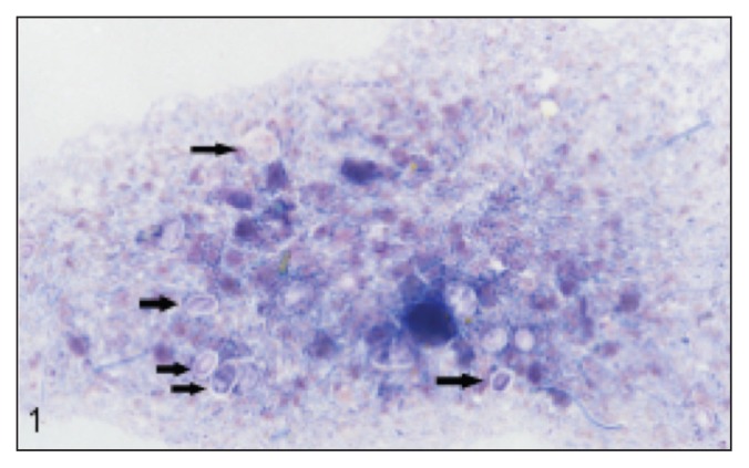
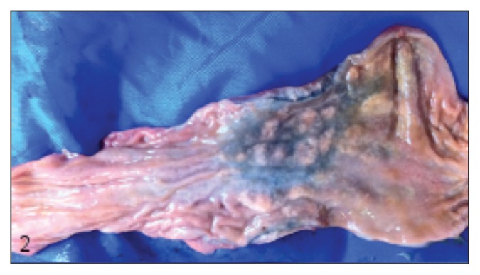
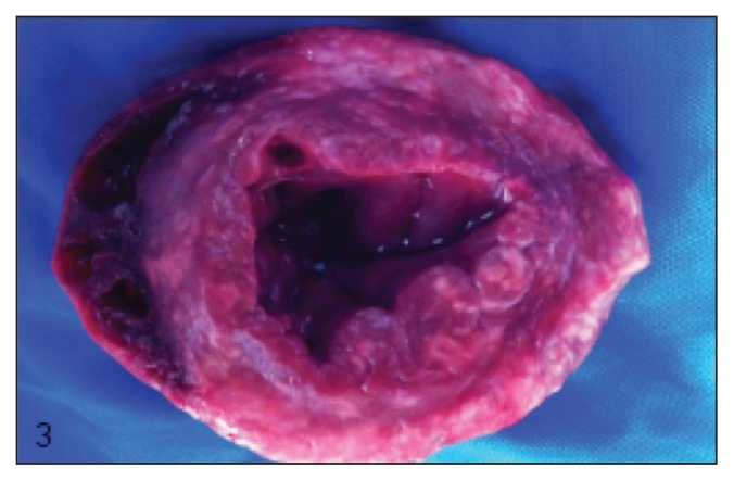
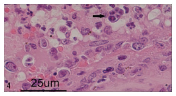
A case of a disseminated algal infection is reported in a young rough-coated collie dog with progressive neurologic deficits, blindness, and hemorrhagic diarrhea. Prototheca zopfii organisms were cultured from feces, urine, and blood. At necropsy, granulomas containing typical organisms were identified within the proximal colon, heart, kidneys, and eyes.
Protothécose chez un chien. Un cas d’infection algoïde est signalé chez un jeune chien Collie à poil court avec des troubles neurologiques progressifs, de la cécité et de la diarrhée hémorragique. Des organismes de type Prototheca zopfii ont été cultivés à partir des fèces, de l’urine et du sang. À la nécropsie, des granulomes contenant des organismes typiques ont été identifiés dans le côlon proximal, le cœur, les reins et les yeux.(Traduit par Isabelle Vallières).
Figures
References
-
- Rubin LF, Lynch RK, Stockman WS. Clinical estimation of lacrimal function in dogs. J Am Vet Med Assoc. 1965;147:946–947. - PubMed
-
- Gelatt KN, Peiffer RL, Jr, Erickson JL, Gum GG. Evaluation of tear formation in the dog, using a modification of the schirmer tear test. J Am Vet Med Assoc. 1975;166:368–370. - PubMed
-
- Knollinger AM, La Croix NC, Barrett PM, Miller PE. Evaluation of a rebound tonometer for measuring intraocular pressure in dogs and horses. J Am Vet Med Assoc. 2005;227:244–248. - PubMed
-
- Hollingsworth SR. Canine protothecosis. Vet Clin North Am Small Anim Pract. 2000;30:1091–1101. - PubMed
-
- Blogg JR, Sykes JE. Sudden blindness associated with protothecosis in a dog. Aust Vet J. 1995;72:147–149. - PubMed
Publication types
MeSH terms
LinkOut - more resources
Full Text Sources