. 2014 Oct 6:12:e0174.
doi: 10.1199/tab.0174. eCollection 2014.

The ubiquitination machinery of the ubiquitin system

Affiliations

The ubiquitination machinery of the ubiquitin system

Judy Callis. Arabidopsis Book. .

Abstract

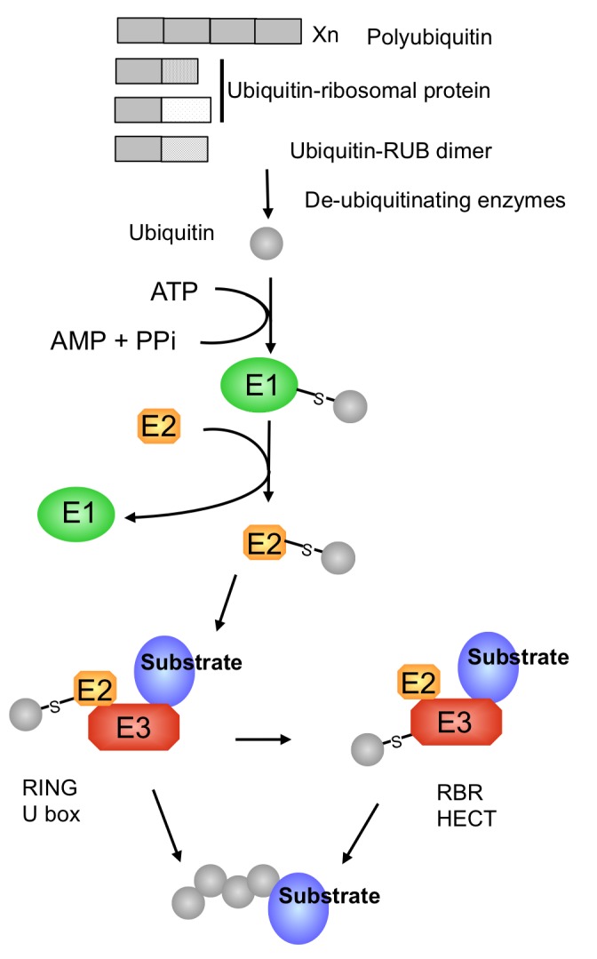
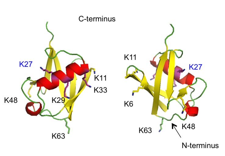
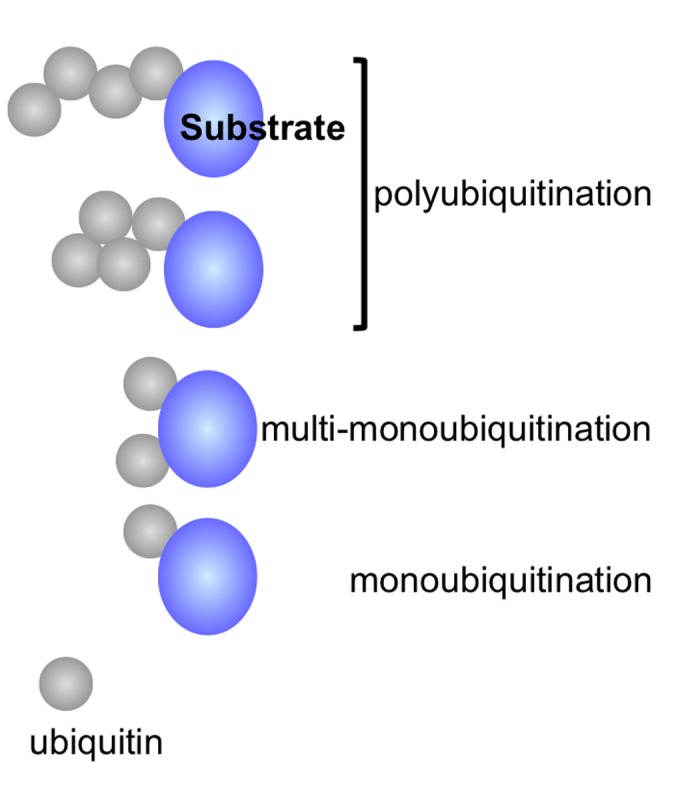
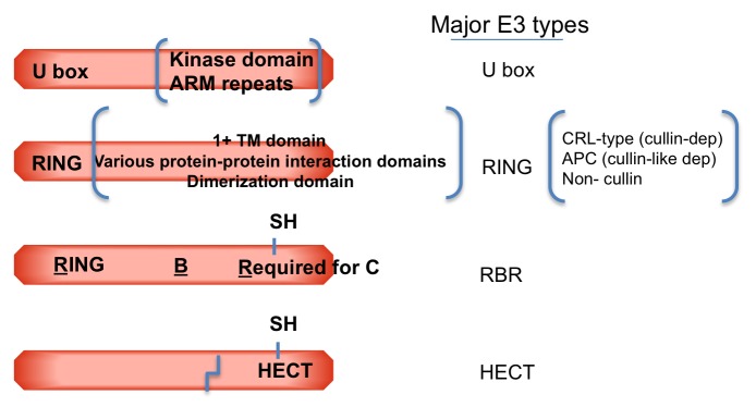
The protein ubiquitin is a covalent modifier of proteins, including itself. The ubiquitin system encompasses the enzymes required for catalysing attachment of ubiquitin to substrates as well as proteins that bind to ubiquitinated proteins leading them to their final fate. Also included are activities that remove ubiquitin independent of, or in concert with, proteolysis of the substrate, either by the proteasome or proteases in the vacuole. In addition to ubiquitin encoded by a family of fusion proteins, there are proteins with ubiquitin-like domains, likely forming ubiquitin's β-grasp fold, but incapable of covalent modification. However, they serve as protein-protein interaction platforms within the ubiquitin system. Multi-gene families encode all of these types of activities. Within the ubiquitination machinery "half" of the ubiquitin system are redundant, partially redundant, and unique components affecting diverse developmental and environmental responses in plants. Notably, multiple aspects of biotic and abiotic stress responses require, or are modulated by, ubiquitination. Finally, aspects of the ubiquitin system have broad utility: as components to enhance gene expression or to regulate protein abundance. This review focuses on the ubiquitination machinery: ubiquitin, unique aspects about the synthesis of ubiquitin and organization of its gene family, ubiquitin activating enzymes (E1), ubiquitin conjugating enzymes (E2) and ubiquitin ligases, or E3s. Given the large number of E3s in Arabidopsis this review covers the U box, HECT and RING type E3s, with the exception of the cullin-based E3s.


Figures

Figure 1.
Figure 1.
Ubiquitin genes and ubiquitination pathway. Ubiquitin is encoded by a family of protein fusions that must be processed by de-ubiquitinating enzymes to release active ubiquitin. Ubiquitin is activated by E1, and thioester conjugated first to E1, then to E2. E2∼Ub interacts with an E3. In the case of RING and U box E3s, an intermediate complex of substrate, E3 and E2∼Ub is required for transfer to substrate. For RBR and HECT-type E3s, E2∼Ub interacts and transfers ubiquitin to an E3 cysteinyl sulfhydryl prior to ubiquitin transfer to substrate.
Figure 2.
Figure 2.
Ribbon diagram representations of human ubiquitin (1.UBQ.pdb). The side chains of the seven lysine residues (6, 11, 27, 29, 33, 48 and 63) are shown in stick form, but not all are visible in each view (180° difference). K27 is in blue type because it was not identified as a ubiquitin-ubiquitin linkage site in Arabidopsis. The C-terminus is at top, the N-terminus at the bottom only visible in the right view. In red, helical regions; in yellow, β-strands.
Figure 3.
Figure 3.
Representation of the diversity of ubiquitinated products. In Arabidopsis, 6/7 lysine residues of ubiquitin serve as ubiquitin attachment sites, forming distinct polyubiquitin chains. K48, K11 and K63 chains are structurally distinct (different chain topology is represented by the top two polyubiquitinated substrates). In addition, substrates can be monoubiquitinated (bottom) or monoubiquitinated at multiple substrate sites (multi-monoubiquitination, second from bottom).
Figure 4.
Figure 4.
Diagrammatic representation of E3 types. From top, U box-, RING-, RBR-, HECT-type E3s contain their respective characteristic domains. RING, for Really Interesting New Gene; HECT, for Homology to E6-AP Carboxy Terminus. The RBR type E3 initially referred to the term RING1-ln Between RING- RING2. More recently, a change in nomenclature (but not abbreviation) was suggested to more accurately represent the biochemical activities of the domains: with RBR representing an abbreviation for: RING- Benign catalytic-Required for catalysis. E2∼Ub interacts with U box and RING domains and a region in HECT E3s upstream of the catalytic SH. The —SH of RBR- and HECT-type E3s is the site of ubiquitin thioester present as an intermediate in ubiquitination of substrates. Many, but not all, U box proteins have kinase or multiple ARM repeats. RING proteins are more diverse; subsets contain 1 or more transmembrane domains (TM), other characterized protein-protein interaction domains and/or other regions implicated in homo-or hetero-oligomerization. The bent line in HECT-type E3 indicates that these proteins are typically very large. The RING and U box domains may be located near the N-terminus, internally or at the C-terminus; the HECT domain is typically at the C-terminus. RING-type E3s include the multiple subunit CRL (for cullin-RING-ligase) types consisting of a RBX (RING BOX) type RING protein, a scaffold cullin protein (one of 3 types in Arabidopsis) and one or more substrate specificity subunits. The APC (anaphase promoting complex) E3 contains a RING protein (APC11) and a cullin-like protein in addition to other subunits.
Figure 5.
Figure 5.
Diagram of the Arabidopsis N-end rule pathway. Top, Proteins co-synthesized with N-terminal ubiquitin coding region (ubiquitin fusion) are cleaved by de-ubiquitinating enzymes to generate a test protein with a specified encoded N-terminal residue (X). The stability of the test protein is determined by the nature of this N-termlnal residue (X). Some amino acids are recognized directly by E3s and are considered 1° de-stabilizing residues. Others require conversion to 1° destabilizing residues. For the 3° de-stabilizing amino acids, glutamine (Q) and asparagine (N), the R amide groups are hydrolyzed by deamidases to generate the 2° de-stabilizing amino acids, glutamate (E) and aspartate (D). An arginine residue is transferred to the N-termini of E and D, converting them to a 1° destabilizing residue. Other basic residues, lysine (K) and histidine (H), are recognized by the same type of E3. Hydrophobic residues, such as leucine (L), phenylalanine (F), tyrosine (Y), tryptophan (W) and isoleucine (I) are also 1° destabilizing, but are recognized by different E3s. Cysteine (C) is considered a 3° destabilizing residue because Is requires oxidation before arginylation.

References

    1. Acconcia F., Sigismund S., Polo S. Ubiquitin in trafficking: The network at work. Exp. Cell Res. 2009;315:1610–1618. - PubMed
    1. Aguilar-Hernández V., Aguilar-Henonin L., Guzmán P. Diversity in the architecture of ATLs, a family of plant ubiquitin-ligases, leads to recognition and targeting of substrates in different cellular environments. PLoS ONE. 2011;6:e23934. - PMC - PubMed
    1. Aguilar-Hernández V., Medina J., Aguilar-Henonin L., Guzmán P. Expansion and diversification of BTL RING-H2 ubiquitin ligases in angiosperms: Putative Rabring7/BCA2 orthologs. PLoS ONE. 2013;8:e72729. - PMC - PubMed
    1. Andersen P., Kragelund B.B., Olsen A.N., Larsen F.H., Chua N.H., Poulsen F.M., Skriver K. Structure and biochemical function of a prototypical Arabidopsis U-box domain. J. Biol. Chem. 2004;279:40053–40061. - PubMed
    1. Aung K., Lin S., Wu C., Huang Y., Su C., Chiou T. Pho2, a phosphate overaccumulator, is caused by a non-sense mutation in a microRNA399 target gene. Plant Physiol. 2006;141:1000–1011. - PMC - PubMed