Paraplegia after epidural-general anesthesia in a Morquio patient with moderate thoracic spinal stenosis
- PMID: 25323122
- PMCID: PMC4288980
- DOI: 10.1007/s12630-014-0247-1
Paraplegia after epidural-general anesthesia in a Morquio patient with moderate thoracic spinal stenosis
Abstract
Purpose: We describe an instance in which complete paraplegia was evident immediately postoperatively after apparently uneventful lumbar epidural-general anesthesia in a patient with Morquio Type A syndrome (Morquio A) with moderate thoracic spinal stenosis.
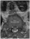
Clinical features: A 16-yr-old male with Morquio A received lumbar epidural-general anesthesia for bilateral distal femoral osteotomies. Preoperative imaging had revealed a stable cervical spine and moderate thoracic spinal stenosis with a mild degree of spinal cord compression. Systolic blood pressure (BP) was maintained within 20% of the pre-anesthetic baseline value. The patient sustained a severe thoracic spinal cord infarction. The epidural anesthetic contributed to considerable delay in the recognition of the diagnosis of paraplegia.
Conclusion: This experience leads us to suggest that, in patients with Morquio A, it may be prudent to avoid the use of epidural anesthesia without very firm indication, to support BP at or near baseline levels in the presence of even moderate spinal stenosis, and to avoid flexion or extension of the spinal column in intraoperative positioning. If the spinal cord/column status is unknown or if the patient is known to have any degree of spinal stenosis, we suggest that the same rigorous BP support practices that are typically applied in other patients with severe spinal stenosis, especially stenosis with myelomalacia, should apply to patients with Morquio A and that spinal cord neurophysiological monitoring should be employed. In the event that cord imaging is not available, e.g., emergency procedures, it would be prudent to assume the presence of spinal stenosis.
Conflict of interest statement
Figures


References
-
- Yasuda E, Fushimi K, Suzuki Y, et al. Pathogenesis of Morquio A syndrome: an autopsied case reveals systemic storage disorder. Mol Genet Metab. 2013;109:301–11. - PubMed
-
- Tomatsu S, Montano AM, Oikawa H, et al. Mucopolysaccharidosis type IVA (Morquio A disease): clinical review and current treatment. Curr Pharm Biotechnol. 2011;12:931–45. - PubMed
Publication types
MeSH terms
Grants and funding
LinkOut - more resources
Full Text Sources
Other Literature Sources
Medical

