. 2014 Oct 21;64(16):1681-94.
doi: 10.1016/j.jacc.2014.06.1204.

Cultured human bone marrow-derived CD31(+) cells are effective for cardiac and vascular repair through enhanced angiogenic, adhesion, and anti-inflammatory effects

Affiliations

Cultured human bone marrow-derived CD31(+) cells are effective for cardiac and vascular repair through enhanced angiogenic, adhesion, and anti-inflammatory effects

Sung-Whan Kim et al. J Am Coll Cardiol. .

Abstract

Background: Cell therapy for cardiovascular disease has been limited by low engraftment of administered cells and modest therapeutic effects. Bone marrow (BM) -derived CD31(+) cells are a promising cell source owing to their high angiovasculogenic and paracrine activities.

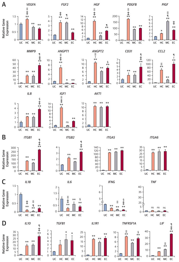
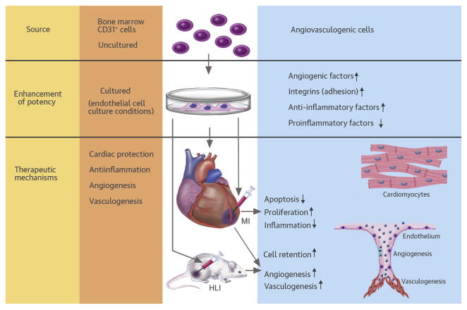
Objectives: This study sought to identify culture conditions that could augment the cell adhesion, angiogenic, and anti-inflammatory activities of BM-derived CD31(+) cells, and to determine whether these cultured CD31(+) cells are effective for cardiac and vascular repair.

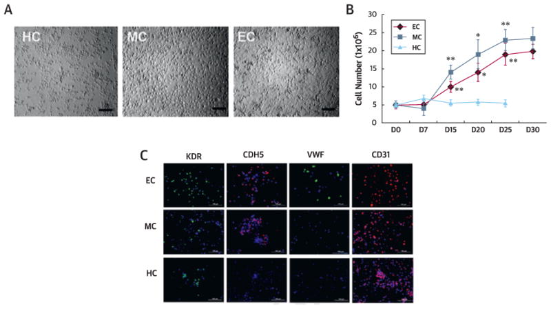
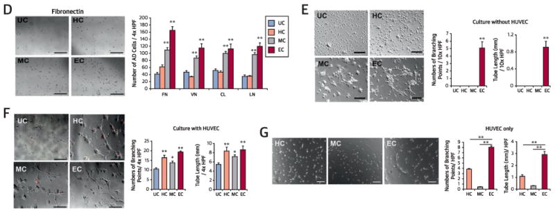
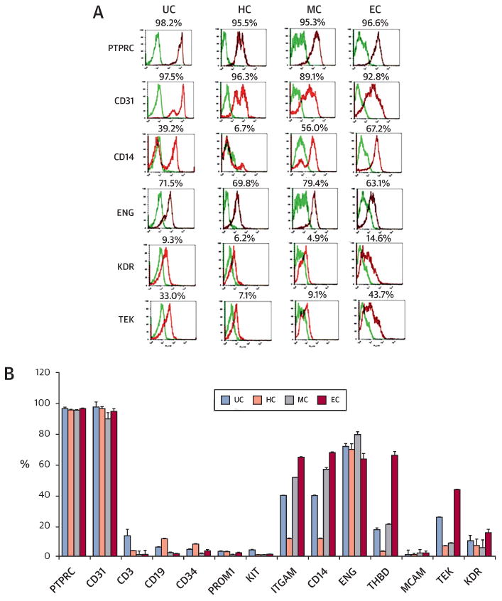
Methods: CD31(+) cells were isolated from human BM by magnetic-activated cell sorting and cultured for 10 days under hematopoietic stem cell, mesenchymal stem cell, or endothelial cell culture conditions. These cells were characterized by adhesion, angiogenesis, and inflammatory assays. The best of the cultured cells were implanted into myocardial infarction (MI) and hindlimb ischemia (HLI) models to determine therapeutic effects and underlying mechanisms.

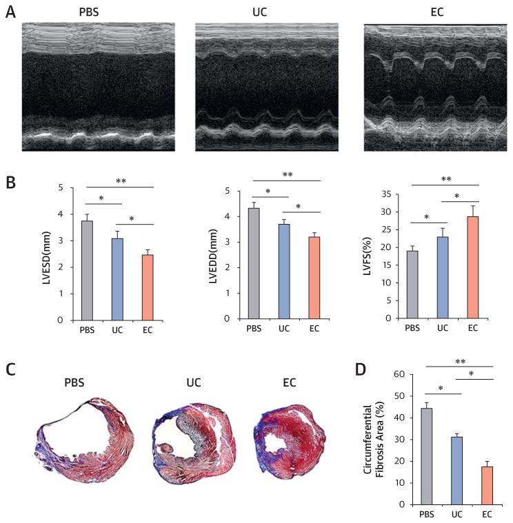
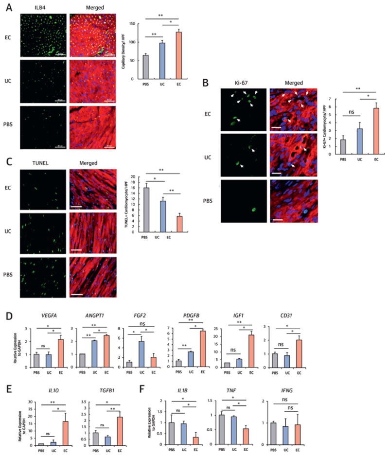
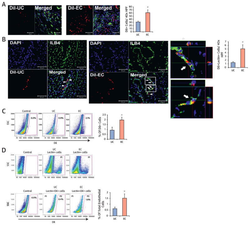
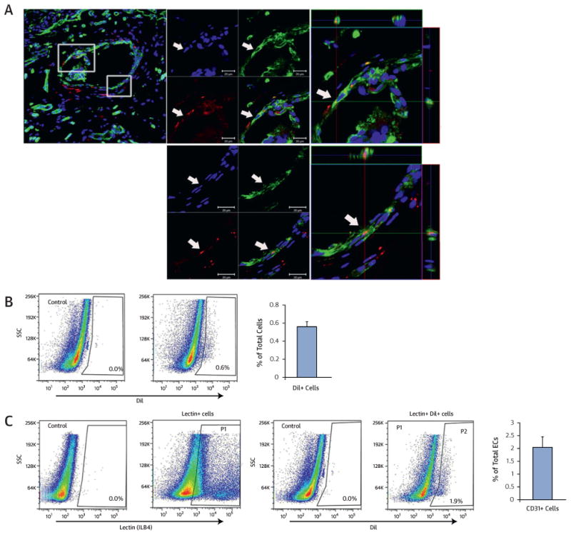
Results: The CD31(+) cells cultured in endothelial cell medium (EC-CD31(+) cells) showed the highest adhesion and angiogenic activities and lowest inflammatory properties in vitro compared with uncultured or other cultured CD31(+) cells. When implanted into mouse MI or HLI models, EC-CD31(+) cells improved cardiac function and repaired limb ischemia to a greater extent than uncultured CD31(+) cells. Histologically, injected EC-CD31(+) cells exhibited higher retention, neovascularization, and cardiomyocyte proliferation. Importantly, cell retention and endothelial transdifferentiation was sustained up to 1 year.

Conclusions: Short-term cultured EC-CD31(+) cells have higher cell engraftment, vessel-formation, cardiomyocyte proliferation, and anti-inflammatory potential, are highly effective for both cardiac and peripheral vascular repair, and enhance survival of mice with heart failure. These cultured CD31(+) cells may be a promising source for treating ischemic cardiovascular diseases.

Keywords: CD31; angiogenesis; engraftment; inflammation; myocardial infarction; peripheral vascular disease.


Figures

Figure 1
Figure 1. In Vitro Cell Biological Characteristics of Cultured and Uncultured CD31+ Cells
(A) Morphologies of CD31+ cells cultured under hematopoietic stem cell (HC), mesenchymal stem cell (MC), and endothelial cell (EC) conditions at day 10. Bars = 200 µm. (B) Growth curves of HC, MC, and EC. n = 5 per group. **p < 0.01, *p < 0.05 vs. HC. (C) Endothelial and mesenchymal culture conditions induced CD31+ cells to express endothelial proteins kinase insert domain receptor (KDR), CDH5, von Willebrand factor (VWF), and CD31 in culture. (D) Cell adhesion assays showed higher adhesion of EC and MC to all tested extracellular matrix proteins (fibronectin [FN], vitronectin [VN], type I collagen [CL], laminin [LN]) compared with uncultured cells (UC). n = 5 per group. Bars = 500 µm. (E) Tube formation potential. Tube lengths and the number of branching points were measured 24 h after seeding of cultured or uncultured CD31+ cells in Matrigel coated plates. n = 5 per group. Bars: 500 µm. (F) Tube formation by coculture with human umbilical vein endothelial cells (HUVEC). Tube lengths and the number of branching points were measured 12 hours after seeding of 1,1∼-dioctadecyl-3,3,3∼,3∼-tetramethylindocarbocyanine perchlorate-labeled cultured or uncultured CD31+ cells in Matrigel-coated plates. n = 5 per group. Bars: 500 µm. (G) Tube formation assay using HUVEC only. Tube lengths and the number of branching points were measured 5 hours after seeding of HUVEC in Matrigel-coated plates. n = 5 per group. **p < 0.01. Bars = 200 µim. (D-F) **p < 0.01 vs. UC, *p < 0.05 vs. UC. AD = adherent; HPF = high-powered field; UC = uncoated.
Figure 2
Figure 2. Flow Cytometric Analyses of Cultured and Uncultured CD31+ Cells
(A) Representative histograms of flow cytometry data. The CD31+ cells cultured under the 3 conditions for 10 days were labeled with phycoerythrin-conjugated (red spectra) or fluorescein isothiocyanate-conjugated (green spectra) antibodies and analyzed by flow cytometry for hematopoietic and endothelial markers. (B) Quantitative analyses (n = 4 each). ENG = endoglin; ITGAM = integrin alpha M; KIT = proto-oncogene c-Kit; MCAM = melanoma cell adhesion molecule; PROM1 = prominin 1; PTPRC = protein tyrosine phosphatase, receptor type, C; TEK = tyrosine kinase, endothelial; THBD = thrombomodulin. Other abbreviations as in Figure 1.
Figure 3
Figure 3. Expression of Angiogenic, Integrin, and Inflammatory Factors
Quantitative reverse transcriptase polymerase chain reaction was performed to measure the level of gene expression from cultured and uncultured CD31+ cells. Various angiogenic (A), cell adhesion (integrin) (B), proinflammatory (C), and anti-inflammatory (D) factors were up-regulated or down-regulated in the cultured groups compared with the uncultured group (n = 4 each; *p < 0.05; **p < 0.01). Individual values were normalized to glyceraldehyde 3-phosphate dehydrogenase (GAPDH) (+p < 0.05, HC vs. EC; p < 0.01, HC vs. EC; §p < 0.05, EC vs. MC; p < 0.01, EC vs. MC; p < 0.01, MC vs. HC). AKT1 = Ak strain transforming oncogene; ANGPT1/2 = angiopoietin 1/2;CCL2 = chemokine (C-C motif) ligand 2; FGF2 = basic fibroblast growth factor; HGF = hepatocyte growth factor; IFNG = interferon gamma; IGF1 = insulin-like growth factor 1; IL(n) = interleukin (n); IL1R1 = interleukin 1 receptor, type1; ITGA5/6 = integrin alpha-5/-6; ITGB1/2 = integrin beta-1/-2; LIF = leukemia inhibitory factor; MMP9 = matrix metallopeptidase 9; PDGFB = platelet-derived growth factor beta; PlGF = placental growth factor; TGFB1 = transforming growth factor beta-1; TNF = tumor necrosis factor; TNFRSF1A = tumor necrosis factor receptor 1A; VEGFA = vascular endothelial growth factor A. Other abbreviations as in Figure 1.
Figure 4
Figure 4. Endothelial Cell Transplantation Is Effective for Infarct Repair
(A) Representative M-mode echocardiograms. (B) Left ventricular end-diastolic dimension (LVEDD) and left ventricular end-systolic dimension (LVESD) were lowest in the endothelial cell (EC) group. Left ventricular fractional shortening (LVFS) was better in EC-injected hearts compared with uncultured cell (UC)-injected or phosphate-buffered saline (PBS)-injected hearts at 3 weeks after myocardial infarction (n = 7 each; *p < 0.05). (C) Representative cross-sectional images of hearts stained with Masson’s trichrome at 4 weeks after cell transplantation. The blue color represents fibrosis. (D) EC transplantation offered the highest reduction in fibrosis area (n = 7; *p < 0.05; **p < 0.01).
Figure 5
Figure 5. Endothelial Cell Transplantation Increased Neovascularization and Cardiomyocyte Proliferation and Protection, and Suppressed Inflammation In Vivo
(A) Isolectin B4 (ILB4) staining for the peri-infarct regions and quantitative analysis of capillary density (n = 7 each; *p < 0.05, **p < 0.01). Bars = 50 μm; sarcomeric α-actinin staining (red). (B) Double immunohistochemistry with Ki-67 and sarcomeric α-actinin antibodies for the peri-infarct regions and quantitative analysis (n = 7 each; *p < 0.05, **p < 0.01). Arrows indicate Ki-67-positive cardiomyocytes. Bars = 20 μm. (C) Terminal deoxynucleotidyl transferase dUTP nick end labeling (TUNEL) assay with sarcomeric α-actinin staining for the peri-infarct regions and quantitative analysis (n = 7 each; *p < 0.05, **p < 0.01). Bars = 50µm. (D-E) Quantitative reverse transcriptase polymerase chain reaction analyses with peri-infarct tissues injected with PBS, UC, or EC. EC transplantation showed highest expression of most angiogenic factors (D) and anti-inflammatory factors (E) and lowest expression of pro-inflammatory factors (F). Data are presented as fold differences from the PBS group (n = 5 each; *p < 0.05; **p < 0.01). Other abbreviations as in Figures 1, 3, and 4.
Figure 6
Figure 6. Cultured CD31+ Cells Showed Higher Engraftment and Endothelial Transdifferentiation Capacity in the Ischemic Heart
(A-B) Histological analyses of engrafted cells in the heart sections 4 weeks after EC or UC (1,1∼-dioctadecyl-3,3,3∼,3∼-tetramethylindocarbocyanine perchlorate [Dil]-labeled, red fluorescence) injection. (A) EC had higher retention compared with UC. n = 5 each. *p < 0.05. (B) Confocal microscopic examination demonstrated that injected UC-CD31+ cells (upper panel) or EC-CD31+ cells (lower panel) were colocalized with ILB4 (white arrows) using 3-dimensional z-stacked orthogonal and mul-tipanel images. Arrows indicate colocalized cells and arrowheads, engrafted cells in the perivascular areas. Quantifications of Dil and ILB4-double positive cells are shown on the right. n = 5 each. *p < 0.05. (C-D) Flow cytometry analyses of digested cardiac tissues at 4 weeks. (C) Higher engraftment of EC compared with UC occurred, n = 5 each. *p < 0.05. (D) Quantification of Dil and ILB4 double-positive cells showing a higher rate of endothelial transdifferentiation in EC compared with UC-injected hearts. n = 5 each. *p < 0.05. P1 gating represents ILB4+ fraction and P2 gating double-positive population for Dil and ILB4. DAPI = 4∼,6-diamidino-2-phenylindole. Other abbreviations as in Figures 1 and 5.
Figure 7
Figure 7. Engraftment and Endothelial Transdifferentiation Capacity at 1 Year in the Ischemic Heart
(A) Cardiac tissues were harvested at 1 year after injection with Dil-labeled EC. Confocal microscopic examination showed perivascular localization of injected EC as well as colocalized Dil-labeled EC (red) with ILB4-stained endothelial cells (green) in vessels in z-stacked orthogonal and multipanel images. Blue, DAPI. (B) Flow cytometry analysis showing presence of Dil-labeled EC in hearts at 1 year after EC injection (n = 5 each). Heart tissues were harvested at 1 year and digested with an enzyme cocktail. (C) Flow cytometry analysis demonstrating Dil and ILB4 double-positive cells showing endothelial transdifferentiation of injected Dil-labeled EC. P1 gating represents the ILB4+ fraction and P2 gating represents the cell population double positive for both Dil and ILB4. Other abbreviations as in Figures 1, 5, and 6.
Central Illustration
Central Illustration. Enhanced therapeutic effects of BM-derived CD31+ cells for cardiac and vascular repair
The bone marrow-derived CD31+ cells cultured in endothelial cell medium (EC-CD31+ cells) showed higher adhesion and angiogenic activities and low inflammatory properties in vitro compared with uncultured or other cultured CD31+ cells. When implanted into mouse myocardial infarction (MI) or hindlimb ischemia (HLI) models, EC-CD31+ cells improved cardiac function and repaired limb ischemia to a greater extent than uncultured CD31+ cells. Mechanistically, injected EC-CD31+ cells induced higher retention, neovascularization, cardiac protection, and cardiomyocyte proliferation, and lower inflammatory reactions.

Comment in

References

    1. Assmus B, Schachinger V, Teupe C, et al. Transplantation of Progenitor Cells and Regeneration Enhancement in Acute Myocardial Infarction (TOPCARE-AMI) Circulation. 2002;106:3009–17. - PubMed
    1. Amado LC, Saliaris AP, Schuleri KH, et al. Cardiac repair with intramyocardial injection of allogeneic mesenchymal stem cells after myocardial infarction. Proc Natl Acad Sci U S A. 2005;102:11474–9. - PMC - PubMed
    1. Yoon YS, Wecker A, Heyd L, et al. Clonally expanded novel multipotent stem cells from human bone marrow regenerate myocardium after myocardial infarction. J Clin Invest. 2005;115:326–38. - PMC - PubMed
    1. Kinnaird T, Stabile E, Burnett MS, et al. Local delivery of marrow-derived stromal cells augments collateral perfusion through paracrine mechanisms. Circulation. 2004;109:1543–9. - PubMed
    1. Rehman J, Li J, Orschell CM, March KL. Peripheral blood “endothelial progenitor cells” are derived from monocyte/macrophages and secrete angiogenic growth factors. Circulation. 2003;107:1164–9. - PubMed

Publication types

MeSH terms

Substances