. 2015 May;22(5):731-42.
doi: 10.1038/cdd.2014.164. Epub 2014 Oct 17.

A rise in NAD precursor nicotinamide mononucleotide (NMN) after injury promotes axon degeneration

Affiliations

A rise in NAD precursor nicotinamide mononucleotide (NMN) after injury promotes axon degeneration

M Di Stefano et al. Cell Death Differ. 2015 May.

Abstract

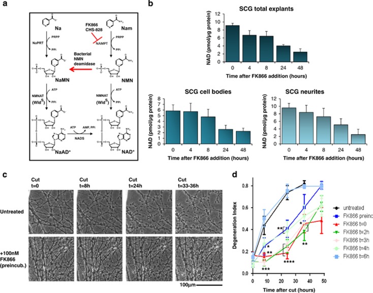
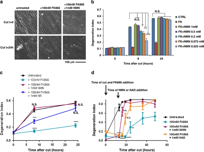
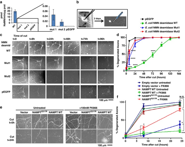
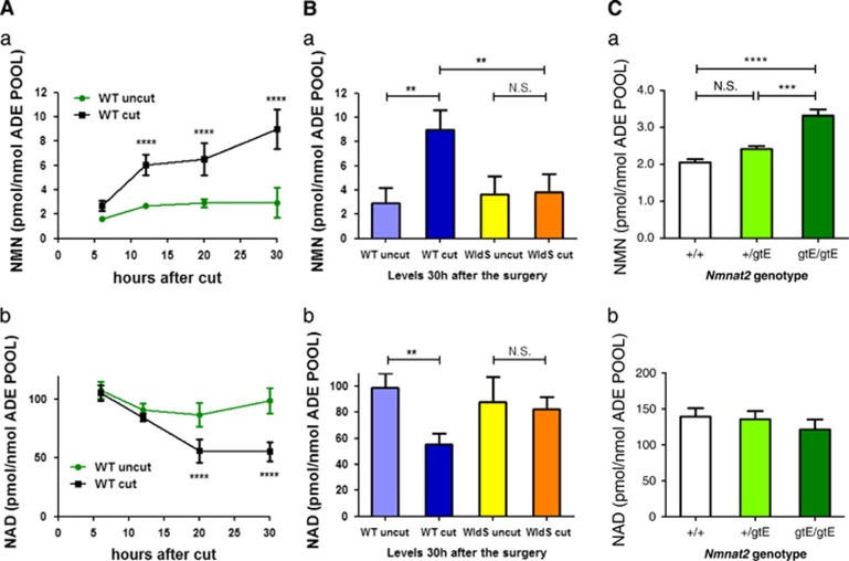
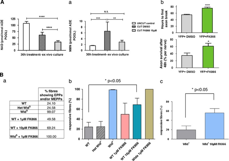
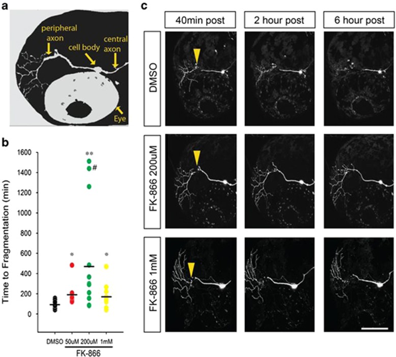
NAD metabolism regulates diverse biological processes, including ageing, circadian rhythm and axon survival. Axons depend on the activity of the central enzyme in NAD biosynthesis, nicotinamide mononucleotide adenylyltransferase 2 (NMNAT2), for their maintenance and degenerate rapidly when this activity is lost. However, whether axon survival is regulated by the supply of NAD or by another action of this enzyme remains unclear. Here we show that the nucleotide precursor of NAD, nicotinamide mononucleotide (NMN), accumulates after nerve injury and promotes axon degeneration. Inhibitors of NMN-synthesising enzyme NAMPT confer robust morphological and functional protection of injured axons and synapses despite lowering NAD. Exogenous NMN abolishes this protection, suggesting that NMN accumulation within axons after NMNAT2 degradation could promote degeneration. Ectopic expression of NMN deamidase, a bacterial NMN-scavenging enzyme, prolongs survival of injured axons, providing genetic evidence to support such a mechanism. NMN rises prior to degeneration and both the NAMPT inhibitor FK866 and the axon protective protein Wld(S) prevent this rise. These data indicate that the mechanism by which NMNAT and the related Wld(S) protein promote axon survival is by limiting NMN accumulation. They indicate a novel physiological function for NMN in mammals and reveal an unexpected link between new strategies for cancer chemotherapy and the treatment of axonopathies.


Figures

Figure 1
Figure 1
FK866 acts within axons to delay degeneration after injury. (a) The salvage pathway of NAD biosynthesis from nicotinamide (Nam) and nicotinic acid (Na). Only NAD biosynthesis from Nam is sensitive to FK866, which potently inhibits NAMPT while having no effect on nicotinic acid phosphoribosyltransferase (NaPRT). The reaction catalysed by bacterial NMN deamidase is also shown. (b) SCG explants were treated with 100 nM FK866 for the indicated times, and then the whole explants (top panel) or the cell bodies (bottom left panel) and neurite fractions (bottom right panel) were separately collected. NAD was determined with an HPLC-based method (see Materials and Methods; n=3, mean and S.D. shown). (c) SCG neurites untreated (top panels) or treated with 100 nM FK866 the day before transection (bottom panels) and imaged after transection at the indicated time points. (d) SCG explants were treated with 100 nM FK866 1 day before or at the indicated times after cutting their neurites. Degeneration index was calculated from three fields in 2–4 independent experiments. The effect of treatment is highly significant when the drug is preincubated or added at 0–4 h after cut (mean ±S.E.M., n=6–12, one-way ANOVA followed by Bonferroni's post-hoc test, *P<0.05, **P<0.01, ***P<0.001, ****P<0.0001, compared with untreated)
Figure 2
Figure 2
NMN promotes Wallerian degeneration. (a) SCG neurites were cut and then left untreated or treated with 100 nM FK866 or with 100 nM FK866 and 1 mM NMN as shown and imaged at the indicated time points. (b) Degeneration index of SCG neurites treated as in panel (a), or with lower NMN concentrations, were calculated from three fields in three independent experiments (n=9, mean±S.E.M., one-way ANOVA with Bonferroni's post-hoc test, *P<0.05, ***P<0.001, ****P<0.0001, compared with untreated at the same time point). (c) Degeneration index of SCG neurites cut and then left untreated or treated with 100 nM FK866, 100 nM FK866 and 1 mM NMN or 100 nM FK866 and 1 mM NR as indicated and imaged at 0, 8 and 24 h were calculated in three fields of three independent experiments (n=9, mean±S.E.M., one-way ANOVA with Bonferroni's post-hoc test, ***P<0.001, ****P<0.0001, compared with untreated at the same time point). (d) Degeneration index of SCG neurites cut and then left untreated or treated as indicated was calculated in three fields of three (for NMN effect) or two (for NAD effect) independent experiments (n= 6–9, mean±S.E.M., one-way ANOVA with Bonferroni's post-hoc test, **P<0.01, ****P<0.0001, compared with untreated at the same time point). NS, not significant
Figure 3
Figure 3
Genetic evidence supporting the role of NMN and NAMPT in axon degeneration. (a) Plasmid cDNA constructs encoding E. coli NMN deamidase WT or its virtually enzymatic inactive mutants Mut1 and Mut2 fused to EGFP, or pEGFP vector alone, were transfected in HEK293T cells. Cells were harvested 48 h after transfection, lysed and NMN deamidase activity was measured as described in Materials and Methods. Activity of the enzymatically inactive mutants was barely detectable (left panel), but on expanding the y axis it was clearly detectable with respect to cell lysates transfected with pEGFP vector alone (right panel) (n=3, mean and S.D. shown). (b) Schematic representation of microinjection experiment. Nuclei of dissociated SCGs were microinjected with the plasmid together with pDsRed2-N1 for expression of DsRed2 fluorescent marker to visualize individual transfected neurons. Two days after microinjections, fluorescent neurites were cut and imaged at different time points. (c and d) NMN deamidase strongly delays degeneration of cut SCG neurites. (c) Constructs expressing WT NMN deamidase or mutants with disrupted enzymatic activity fused to EGFP were microinjected into SCG nuclei along with pDsRed2-N1. EGFP fluorescence within the neurites confirmed expression and homogeneous localization of the expressed proteins in cell bodies and axons. SCG neurites were cut 48 h after microinjection, and DsRed2 or EGFP images were acquired at different time points after cut as indicated. (d) The percentage of fluorescent-degenerated axons was calculated in five fields of three independent experiments. (mean ±S.E.M., n=15, one-way ANOVA followed by Bonferroni's post-hoc test *P<0.05, ****P<0.0001). (e and f) Drug-resistant NAMPT restores rapid axon degeneration. (e) Representative images of SCG neurites 0 and 24 h after cutting and microinjected as in panel (b) with empty vector or with vectors expressing drug-resistant or WT NAMPT. SCGs were left untreated or 100 nM FK866 was added just after transection as indicated. (f) The percentage of fluorescent degenerated axons was calculated in 3–6 fields of three independent experiments as in panel (e). The results show that drug-resistant NAMPTG217R expression restores rapid axon degeneration after FK866 treatment partially at 8 h and totally at 24 h after cut (mean±S.E.M., n=6–18, one-way ANOVA followed by Bonferroni's post-hoc test *P<0.05, ***P<0.001). NS, not significant
Figure 4
Figure 4
NMN accumulates in injured sciatic nerves before degeneration. (A) NMN (a) and NAD (b) levels were determined in mouse sciatic nerves at the indicated time points after nerve transection, and in the contralateral uncut nerve, and normalized to the total adenylate pool (ATP+ADP+AMP) as a measure of nucleotide yield (n=4 per time point; mean±S.E.M., two-way ANOVA with Bonferroni's post-hoc test, ****P<0.0001). Minor changes in the unlesioned, contralateral nerves likely reflect that they are not completely isolated from the effects of the operation such as anaesthesia and secondary effects. (B) NMN (a) and NAD (b) levels in sciatic nerves lesioned for 30 h of WT and WldS mice (n=3–4, one-way ANOVA with Bonferroni's post-hoc test, **P<0.01). (C) NMN (a) and NAD (b) levels were determined in the whole brain of WT E18.5 embryos (+/+) and of E18.5 embryos of heterozygous (+/gtE) or homozygous (gtE/gtE) for the Nmnat2gtE allele from which NMNAT2 expression is non-detectable. There is a clear increase in NMN levels in homozygous mice that do not express any NMNAT2 (n=5, one-way ANOVA with Tukey's post-hoc test, ***P<0.001, ****P<0.0001)
Figure 5
Figure 5
FK866 is effective ex vivo and preserves functional axons and neuromuscular synapses. (A) FK866 delays Wallerian degeneration ex vivo. (a) NAD (left panel) and NMN (right panel) levels were determined in mouse sciatic nerve explants cultured for 30 h in the presence of 10 μM FK866 or vehicle (DMSO) and normalized against the total adenylate pool (n=10, mean±S.E.M., unpaired Student's t-test, **P<0.01, ***P<0.001, ****P<0.0001). (b) Rate of axon degeneration in YFP-H nerve explants cultured for 135 h in the presence of 10 μM FK866 or DMSO control. The upper panel shows the time until the first axon break appeared in each nerve. The lower panel shows the percentage of surviving axons in each nerve after 48 h (n=5–10 nerves; mean±S.E.M., one-way ANOVA with Bonferroni's post-hoc test, *P<0.05, ****P<0.0001). (B) FK866 applied to WT muscles mimics synaptic protection conferred by WldS ex vivo. The number of responsive fibres, that is, those showing EPPs upon stimulation and/or spontaneous miniature EPPs, was analysed in isolated tibial nerve/FDB muscle preparations that had been incubated in MPS containing FK866 in panels (a and b) for 16–20 h (n=4–5 muscles in each group) and in panel (c) for 42–48 h (n=8 muscles in both groups). FK866 dose-dependently delayed synaptic degeneration in WT muscles at 16–20 h (P<0.05; ANOVA with post-hoc Dunnett's test) and potentiated the protective effect of WldS at 48 h (P<0.05; t-test). Note that synaptic degeneration in FDB muscles from WldS mice was markedly more rapid ex vivo than in vivo, probably reflecting the shorter nerve stump length and altered environment. However, the phenotype is readily distinguished from WT ex vivo, with incubation in MPS at 32 °C. het=heterozygous
Figure 6
Figure 6
FK866 delays Wallerian degeneration in vivo. (a) Schematic of larval zebrafish head indicating position of trigeminal neurons and axons, relative to the fish eye. (b) Time to beginning of fragmentation following laser axotomy in larvae pretreated for 2 h with vehicle (1% DMSO) (n=25), 50 μM (n=9), 200 μM (n=11) or 1 mM (n=10) FK866. Each circle represents one experiment; horizontal bar denotes average degeneration time (# indicates data from axon still intact >24 h, *P<0.05; **P<0.001). (c) Confocal images of trigeminal neurons postaxotomy labeled with DsRed-Express and treated with 1% DMSO or FK866. Arrowheads point to the site of axotomy. Scale bar, 100 μm

References

    1. Stoll G, Jander S, Myers RR. Degeneration and regeneration of the peripheral nervous system: from Augustus Waller's observations to neuroinflammation. J Peripher Nerv Syst. 2002;7:13–27. - PubMed
    1. Lunn ER, Perry VH, Brown MC, Rosen H, Gordon S. Absence of Wallerian degeneration does not hinder regeneration in peripheral nerve. Eur J Neurosci. 1989;1:27–33. - PubMed
    1. Conforti L, Gilley J, Coleman MP. Wallerian degeneration: an emerging axon death pathway linking injury and disease. Nat Rev Neurosci. 2014;15:394–409. - PubMed
    1. Agostini M, Bedin C, Pucciarelli S, Enzo M, Briarava M, Seraglia R, et al. APC I1307K mutations and forkhead box gene (FOXO1A): another piece of an interesting correlation. Int J Biol Markers. 2012;27:13–19. - PubMed
    1. Martin SM, O'Brien GS, Portera-Cailliau C, Sagasti A. Wallerian degeneration of zebrafish trigeminal axons in the skin is required for regeneration and developmental pruning. Development. 2010;137:3985–3994. - PMC - PubMed

Publication types

MeSH terms

Substances