Review
. 2014 Sep;24(5):510-8.
doi: 10.1111/bpa.12178.

Neuropathology and genetics of cerebroretinal vasculopathies

Affiliations
Review

Neuropathology and genetics of cerebroretinal vasculopathies

Grant R Kolar et al. Brain Pathol. 2014 Sep.

Abstract

Cerebroretinal vasculopathy (CRV) and the related diseases hereditary endotheliopathy with retinopathy, neuropathy, and stroke (HERNS), hereditary vascular retinopathy (HVR) and hereditary systemic angiopathy (HSA) [subsequently combined as retinovasculopathy and cerebral leukodystrophy (RVCL)] are devastating autosomal-dominant disorders of early to middle-age onset presenting with a core constellation of neurologic and ophthalmologic findings. This family of diseases is linked by specific mutations targeting a core region of a gene. Frameshift mutations in the carboxyl-terminus of three prime exonuclease-1 (TREX1), the major mammalian 3' to 5' DNA exonuclease on chromosome 3p21.1-p21.3, result in a systemic vasculopathy that follows an approximately 5-year course leading to death secondary to progressive neurologic decline, with sometimes a more protracted course in HERNS. Neuropathological features include a fibrinoid vascular necrosis or thickened hyalinized vessels associated with white matter ischemia, necrosis and often striking dystrophic calcifications. Ultrastructural studies of the vessel walls often demonstrate unusual multilaminated basement membranes.

Keywords: CRV; HERNS; and stroke; cerebral microvascular disease; hereditary endotheliopathy with retinopathy; hereditary vascular retinopathy; nephropathy; ocular microvascular disease.


Figures

Figure 1
Figure 1
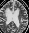
T2 weighted MRI from HERNS patient. MRI demonstrates elevated periventricular signal with enlargement of the lateral ventricles and cortical sulci.
Figure 2
Figure 2
Gross appearance of lesions from CRV and HERNS patients. A. Fixed section demonstrates large space occupying lesion (arrow) in the left frontal lobe white matter and periventricular region. The lesion also involves portions of the deep gray nuclei and obliterates the left lateral ventricle, expands the cortical sulci and causes a midline shift. B. Fixed section from right parietal lobe of the same patient shows a separate periventricular white matter lesion (arrow) with central cores of white, firm parenchyma surrounded by a soft, translucent region consistent with the appearance of an infarct. The gray matter is spared. C. Fixed section from a left half coronal section at the level of the hippocampus from a HERNS patient showing a periventricular lesion (arrow) composed of a soft, translucent lesion with central punctate cores of white, firm parenchyma.
Figure 3
Figure 3
White matter from a CRV patient demonstrating astrocytosis, vascular hyalinization (arrowhead) and an infarct with a central region of dystrophic calcification (arrow).
Figure 4
Figure 4
Ischemic white matter from a CRV patient demonstrates vascular wall hyalinization (arrow) with expanded adventitia. In some vessels, this is accompanied by increased numbers of inflammatory cells but vasculitic damage is not appreciated. A number of smaller vessels within the ischemic white matter are themselves necrotic.
Figure 5
Figure 5
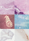
Vascular and white matter lesion in HERNS and CRV. A. A CRV patient demonstrating a moderately sized vessel with fibrinoid vascular necrosis. In the surrounding region, there is an influx of neutrophils consistent with a recently necrotic lesion. B. Reactive astrocytosis is seen in the white matter of a CRV patient adjacent to a region of dystrophic calcifications (seen at lower left). A vascular telangiectasia is also adjacent to the dystrophic calcifications. C. A cluster of vessels from a HERNS patient demonstrate significant hyalinosis of the wall along with adventitial fibrosis. D. A luxol fast blue stain of a region of white matter in a HERNS patient demonstrates focal regions of demyelination that also correspond to areas of neurofilament absence (by IHC), consistent with microscopic ischemic foci. E. Overview of the white and gray matter distribution of CD68 in a HERNS patient demonstrating a patchy, exclusively white matter distribution. F. A magnified view of the same section (as E) demonstrating a population of macrophages within one of the small ischemic foci in the white matter.
Figure 6
Figure 6
Vascular phenotypes seen within the white matter of CRV patients. The morphology of the vasculature in CRV patients differs with proximity to ischemic lesions. Thickening in the vessel walls is common. In some cases, the wall has cellular elements consisting of smooth muscle cells and occasional CD68+ cells (vessels A and B). In these, few CD45+ cells are seen. In other regions, often bordering regions of ischemia (vessel C), the amount of immunostaining of smooth muscle actin and CD68 is decreased. Increased CD45+ cells are seen in some of these vessels. Elsewhere, impressive adventitial thickening can be seen with evidence of loss of lumen integrity (RBC leaking) particularly within regions of ischemia (vessel D). These vessels show no definite cellular elements in their walls.
Figure 7
Figure 7
Vascular ultrastructure of the brain and kidney in CRV patients. Ultrastructure of the vasculature from various regions of the brain (panels A to D) and kidney (panel E and F) of an untreated patient collected within 3 h of death demonstrated striking changes in vascular morphology. A. A region of grossly normal appearing brain demonstrated vessels with normal appearing basement membrane, thin endothelial cells and a patent lumen. The surrounding parenchyma of the brain demonstrated unremarkable neuropil and cellular elements. B. A region bordering an ischemic lesion showed changes to the collagen layers surrounding vessels including increased layers of collagen and a basement membrane that shows thickening and regions with more than one lamina densa layer (examples at arrows). The endothelial cells also demonstrated increased vesicles and had coarse cytoplasm. The surrounding neuropil contains regions with increased lipofuscin and the pericyte foot processes surrounding the vessels have darker, dense cytoplasm. C. A vessel taken within a lesion demonstrates much thicker basement membrane material with entrapped collagen pockets and regions with duplicated lamina densa (arrows). The endothelial cells in this region are far thicker than in grossly normal areas. D. Higher magnification of a vessel wall from the brain showing duplicated lamina densa (stars). A pericyte foot process demonstrates darkened cytoplasm. E. Vessels from the kidney of a different patient show areas with multilaminated basement membranes consisting of alternating lamina densa and lamina lucida. F. A higher magnification of a vessel from the kidney demonstrates multiple layers of lamina densa (stars). Abbreviations: Oligo = oligodendrocyte; Endo = endothelial cell; RBC = erythrocyte; Lipo = lipofuscin; Peri = pericyte; BM = basement membrane.

References

    1. Chowdhury D, Beresford PJ, Zhu P, Zhang D, Sung J‐S, Demple B et al (2006) The exonuclease TREX1 is in the SET complex and acts in concert with NM23‐H1 to degrade DNA during granzyme A‐mediated cell death. Mol Cell 23:133–142. - PubMed
    1. Cohn AC, Kotschet K, Veitch A, Delatycki MB, McCombe MF (2005) Novel ophthalmological features in hereditary endotheliopathy with retinopathy, nephropathy and stroke syndrome. Clin Experiment Ophthalmol 33:181–183. - PubMed
    1. Crow YJ, Hayward BE, Parmar R, Robins P, Leitch A, Ali M et al (2006) Mutations in the gene encoding the 3“‐5” DNA exonuclease TREX1 cause Aicardi‐Goutières syndrome at the AGS1 locus. Nat Genet 38:917–920. - PubMed
    1. Dumitrache LC, Hu L, Hasty P (2009) TREX2 exonuclease defective cells exhibit double‐strand breaks and chromosomal fragments but not Robertsonian translocations. Mutat Res 662:84–87. - PMC - PubMed
    1. Grand MG, Kaine J, Fulling K, Atkinson J, Dowton SB, Farber M et al (1988) Cerebroretinal vasculopathy. A new hereditary syndrome. Ophthalmology 95:649–659. - PubMed

Publication types

MeSH terms

Substances

Supplementary concepts