. 2014 Nov;21(11):472-482.
doi: 10.1038/cgt.2014.53. Epub 2014 Oct 17.

Bortezomib sensitizes non-small cell lung cancer to mesenchymal stromal cell-delivered inducible caspase-9-mediated cytotoxicity

Affiliations

Bortezomib sensitizes non-small cell lung cancer to mesenchymal stromal cell-delivered inducible caspase-9-mediated cytotoxicity

Miki Ando et al. Cancer Gene Ther. 2014 Nov.

Abstract

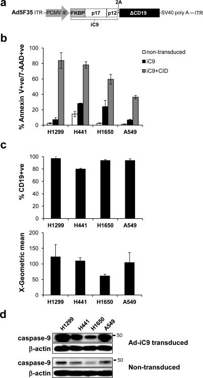
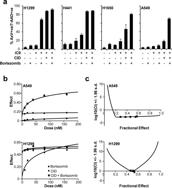
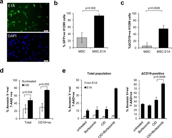
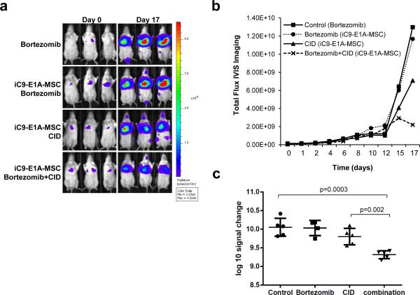
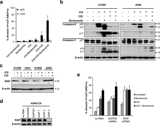
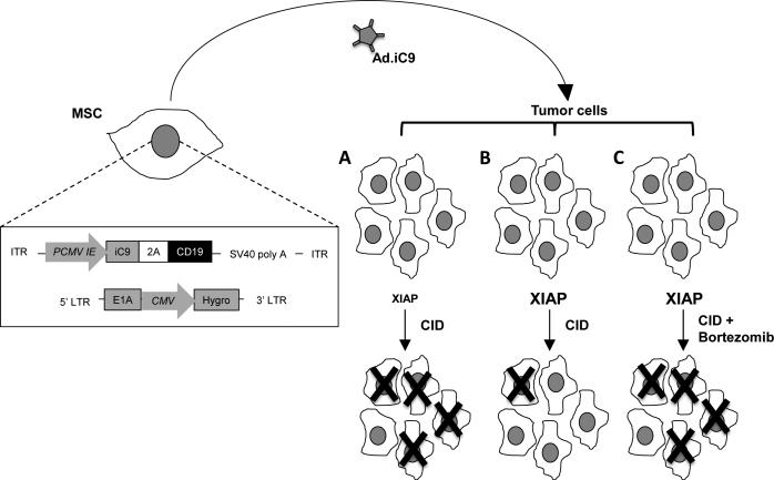
Delivery of suicide genes to solid tumors represents a promising tumor therapy strategy. However, slow or limited killing by suicide genes and ineffective targeting of the tumor has reduced effectiveness. We have adapted a suicide system based on an inducible caspase-9 (iC9) protein that is activated using a specific chemical inducer of dimerization (CID) for adenoviral-based delivery to lung tumors via mesenchymal stromal cells (MSCs). Four independent human non-small cell lung cancer (NSCLC) cell lines were transduced with adenovirus encoding iC9, and all underwent apoptosis when iC9 was activated by adding CID. However, there was a large variation in the percentage of cell killing induced by CID across the different lines. The least responsive cell lines were sensitized to apoptosis by combined inhibition of the proteasome using bortezomib. These results were extended to an in vivo model using human NSCLC xenografts. E1A-expressing MSCs replicated Ad.iC9 and delivered the virus to lung tumors in SCID mice. Treatment with CID resulted in some reduction of tumor growth, but addition of bortezomib led to greater reduction of tumor size. The enhanced apoptosis and anti-tumor effect of combining MSC-delivered Ad.iC9, CID and bortezomib appears to be due to increased stabilization of active caspase-3, as proteasomal inhibition increased the levels of cleaved caspase-9 and caspase-3. Knockdown of X-linked inhibitor of apoptosis protein (XIAP), a caspase inhibitor that targets active caspase-3 to the proteasome, also sensitized iC9-transduced cells to CID, suggesting that blocking the proteasome counteracts XIAP to permit apoptosis. Thus, MSC-based delivery of the iC9 suicide gene to human NSCLC effectively targets lung cancer cells for elimination. Combining this therapy with bortezomib, a drug that is otherwise inactive in this disease, further enhances the anti-tumor activity of this strategy.


Figures

Figure 1
Figure 1
CID induces iC9-dependent apoptosis in transduced NSCLC cell lines. (a) Schematic representation of the adenoviral iC9 bi-cistronic vector. The construct contains the suicide gene iC9 and truncated CD19 (ΔCD19) as a marker separated by a 2A sequence. (b) NSCLC cell lines were transduced with Ad-iC9. 48 hr later cells were left untreated or treated with CID (80nM). Apoptosis was assessed by flow cytometry for Annexin V binding and 7-AAD staining 24 hours after treatment. Error bars represent standard deviation. (c) iC9-transduced cells were assessed for ΔCD19 expression and corresponding MFI by flow cytometry for staining with anti-CD19 antibody 72 hr following transduction. (d) Lysates from cells in (c) and non-transduced cells were blotted for the presence of caspase-9 with an anti-caspase-9 antibody.
Figure 2
Figure 2
Bortezomib enhances CID-induced apoptosis of iC9-transduced cells. (a) Non transduced and iC9-transduced NSCLC cell lines were left untreated or treated with bortezomib (80 nM) in the presence or absence of CID (80 nM) as indicated. Apoptosis was assessed by flow cytometry for Annexin V binding and 7-AAD staining 24 hours after treatment. Error bars represent standard deviation. (b) iC9-transduced A549 and H1299 cells were treated with the indicated amounts of CID, bortezomib or a combination of both. Apoptosis was assessed by flow cytometry for Annexin V binding and 7-AAD staining 24 hours after treatment (see Figure S2). Dose-effect curves were plotted using CalcuSyn™ software. (c) Combination indices were calculated from the data in (b) and plotted using CalcuSyn™ software. CI value less than 1.0 is considered synergistic.
Figure 3
Figure 3
E1A-modified MSC deliver Ad-iC9 to tumor cells that are targeted to undergo apoptosis with CID and bortezomib. (a) MSC were transduced with retrovirus encoding the E1A gene. Five days later cells were fixed and stained with anti-E1A antibody and an Alexa Fluor 488-conjugated secondary antibody (green) and counter-stained with DAPI (blue). Representative images of 1 of 5 donors showing E1A expression in the nuclei of 40-60% of MSC. Scale bars represent 50μm. (b) H1299 cells were incubated with supernatant from Ad.GFP-transduced MSC or Ad.GFP-transduced MSC expressing E1A. GFP expression was measured by flow cytometry 3 days later. Error bars represent standard deviation. (c) H1299 cells were incubated with supernatant from Ad.iC9-transduced MSC or Ad.iC9-transduced MSC expressing E1A. iC9 expression was assessed by flow cytometry for CD19 staining 3 days later. Error bars represent standard deviation. (d) H1299 cells were infected with Ad.iC9 as in (c) followed by treatment with CID (80 nM). Apoptosis was assessed by flow cytometry for Annexin V binding and 7-AAD staining 24 hours after treatment. Error bars represent standard deviation. (e) A549 cells were incubated with supernatant from Ad.iC9-transduced MSC or Ad.iC9-transduced MSC expressing E1A for 3 days. Cells were left untreated or treated with CID (80 nM), bortezomib (80 nM), or both. Apoptosis was assessed by flow cytometry for Annexin V binding and 7-AAD staining 24 hours after treatment (left). Cells incubated with supernatants from E1A positive MSC were gated for ΔCD19 expression and the results are shown (right). Error bars represent standard deviation.
Figure 4
Figure 4
AdiC9-E1A-MSC treatment in combination with bortezomib suppresses tumor growth. (a) SCID-Beige mice were engrafted with FFluc-labeled A549 cells intravenously (IV) followed by IV infusion with iC9-E1A-MSC where indicated. Mice received intraperitoneal injections of CID (50 μg), bortezomib (0.3 mg/kg) or both as described in “Methods”. Tumor growth was monitored by in vivo imaging to measure bioluminescence. Representative images of bioluminescence in mice show the tumor site and tumor size on Day 0 and Day 17. (b) Growth of tumors from mice in each treatment group was measured by assessing mean bioluminescence (5 mice per group). (c) Total tumor growth after 17 days is represented as log10 signal change. Error bars represent standard deviation.
Figure 5
Figure 5
Proteasome inhibition enhances stabilization of active caspase-3. (a) iC9-transduced A549 cells were left untreated or treated with CID (200 nM), bortezomib (80 nM), or a combination of both in the presence or absence of qVD-OPH (20 μM). Apoptosis was assessed by flow cytometry for Annexin V binding and 7-AAD staining 24 hours after treatment. Error bars represent standard deviation. (b) Non-transduced or Ad-iC9-transduced H1299 cells (left) or non-transduced or Ad-iC9-transduced A549 cells (right) were treated with bortezomib (80 nM), CID (200 nM) or both as indicated, and lysates were made 24 hr later. Equal amounts of total protein were analyzed by immunoblotting for caspase-9, cleaved caspase-9, caspase-3, cleaved caspase-3 or actin (as a loading control). (c) NSCLC cell lines or iC9-transduced NSCLC cell lines were left untreated or treated with CID (200 nM) as indicated, and lysates were made 24 hr later. Equal amounts of total protein were analyzed by immunoblotting for XIAP expression or actin (as a loading control). (d) A549 cells were stably transduced with non-targeting (scramble) shRNA, control shRNA (GAPDH) or three different shRNAs targeting XIAP (XIAP1, XIAP2 and XIAP3). Whole cell lysates were analyzed for XIAP knockdown by immunoblot. (e) A549 cells, XIAP knockdown A549 cells (using XIAP3 shRNA) or GAPDH knockdown A549 cells were transduced with Ad.iC9 followed by treatment with bortezomib (80nM), CID (200nM) or both. Apoptosis was assessed by flow cytometry for Annexin V binding and 7-AAD staining 24 hours after treatment. Error bars represent standard deviation.
Figure 6
Figure 6
Proposed model for iC9-E1A-MSC therapy. Schematic representation of MSC modified to express E1A via retroviral transduction and iC9 via adenoviral transduction is shown (left). Infused iC9-E1A-MSC migrate toward the tumor site where they release Ad.iC9, which, in turn, infects lung tumor cells. When tumor cells that have low XIAP activity are infected with Ad.iC9 and exposed to CID, the majority of them will undergo apoptosis (A). When XIAP activity is high, CID will have little effect as XIAP targets active caspases to the proteasome (B). XIAP activity in the latter tumors can be overcome by further treatment with bortezomib to block proteasome-mediated degradation of active caspases, enhancing apoptosis (C).

References

    1. Duarte S, Carle G, Faneca H, de Lima MC, Pierrefite-Carle V. Suicide gene therapy in cancer: where do we stand now? Cancer Lett. 2012;324(2):160–170. - PubMed
    1. Mohit E, Rafati S. Biological delivery approaches for gene therapy: Strategies to potentiate efficacy and enhance specificity. Mol Immunol. 2013;56(4):599–611. - PubMed
    1. Di Stasi A, Tey SK, Dotti G, Fujita Y, Kennedy-Nasser A, Martinez C, et al. Inducible apoptosis as a safety switch for adoptive cell therapy. N Engl J Med. 2011;365(18):1673–1683. - PMC - PubMed
    1. Ramos CA, Asgari Z, Liu E, Yvon E, Heslop HE, Rooney CM, et al. An inducible caspase 9 suicide gene to improve the safety of mesenchymal stromal cell therapies. Stem Cells. 2010;28(6):1107–1115. - PMC - PubMed
    1. Straathof KC, Pule MA, Yotnda P, Dotti G, Vanin EF, Brenner MK, et al. An inducible caspase 9 safety switch for T-cell therapy. Blood. 2005;105(11):4247–4254. - PMC - PubMed

Publication types

MeSH terms