Review
. 2015 Jan 2;116(1):167-82.
doi: 10.1161/CIRCRESAHA.116.303554. Epub 2014 Oct 16.

The mitochondrial dynamism-mitophagy-cell death interactome: multiple roles performed by members of a mitochondrial molecular ensemble

Affiliations
Review

The mitochondrial dynamism-mitophagy-cell death interactome: multiple roles performed by members of a mitochondrial molecular ensemble

Gerald W Dorn 2nd et al. Circ Res. .

Abstract

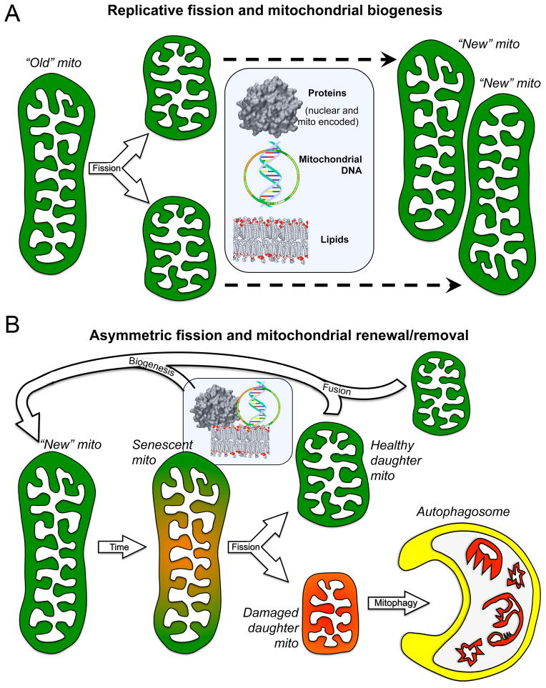
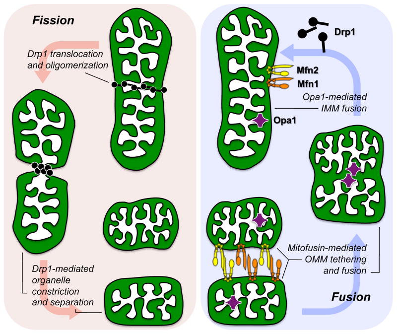
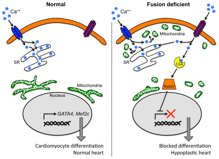
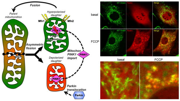
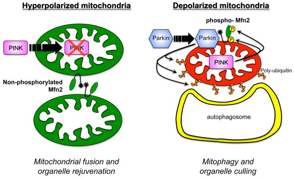
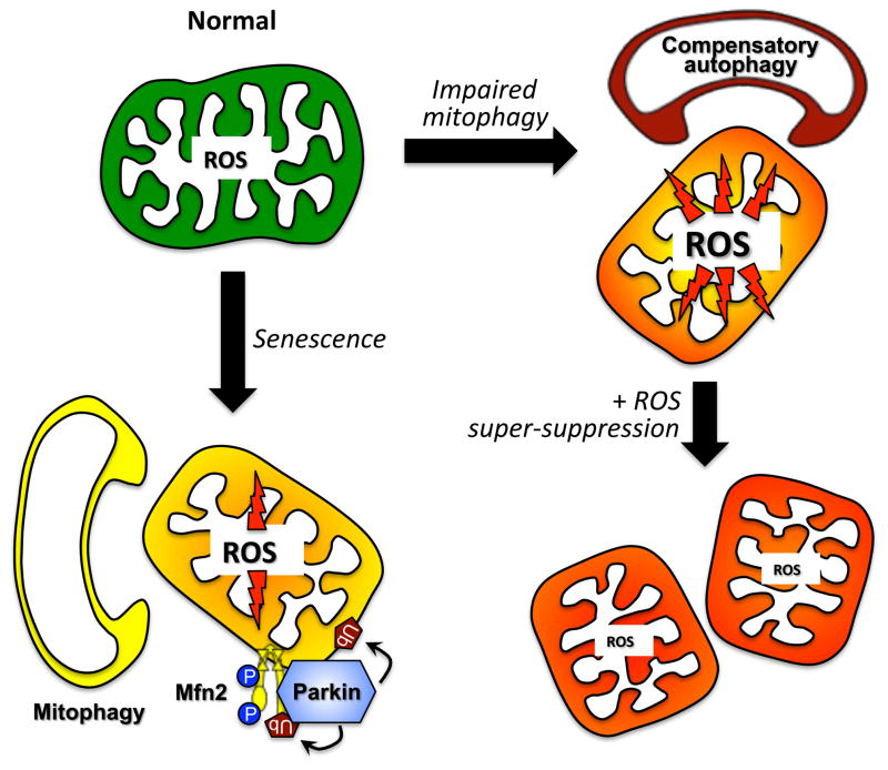
Mitochondrial research is experiencing a renaissance, in part, because of the recognition that these endosymbiotic descendants of primordial protobacteria seem to be pursuing their own biological agendas. Not only is mitochondrial metabolism required to produce most of the biochemical energy that supports their eukaryotic hosts (us) but mitochondria can actively (through apoptosis and programmed necrosis) or passively (through reactive oxygen species toxicity) drive cellular dysfunction or demise. The cellular mitochondrial collective autoregulates its population through biogenic renewal and mitophagic culling; mitochondrial fission and fusion, 2 components of mitochondrial dynamism, are increasingly recognized as playing central roles as orchestrators of these processes. Mitochondrial dynamism is rare in striated muscle cells, so cardiac-specific genetic manipulation of mitochondrial fission and fusion factors has proven useful for revealing noncanonical functions of mitochondrial dynamics proteins. Here, we review newly described functions of mitochondrial fusion/fission proteins in cardiac mitochondrial quality control, cell death, calcium signaling, and cardiac development. A mechanistic conceptual paradigm is proposed in which cell death and selective organelle culling are not distinct processes, but are components of a unified and integrated quality control mechanism that exerts different effects when invoked to different degrees, depending on pathophysiological context. This offers a plausible explanation for seemingly paradoxical expression of mitochondrial dynamics and death factors in cardiomyocytes wherein mitochondrial morphometric remodeling does not normally occur and the ability to recover from cell suicide is severely limited.

Keywords: apoptosis; autophagy; mitochondria; mitochondrial dynamics.


Figures

Figure 1
Figure 1. Consequences of replicative vs asymmetric mitochondrial fission
A. Replicative fission of one healthy “old” parent mitochondrion produces two small healthy daughter organelles that incorporate biogenically produced protein, DNA, and lipids (central rectangle) to grow into “new” mitochondria. B. Asymmetric fission of a damaged or senescent mitochondrion produces one healthy daughter that fuses with other healthy organelles to regenerate the collective, and one severely damaged/depolarized (red) daughter that is rapidly eliminated by autophagosomal engulfment, thereby protecting the cell from mito toxicity and providing new recycled components for biogenic repair.
Figure 2
Figure 2. Structural differences between mouse fibroblast and adult cardiomyocyte mitochondria
A. Top left is Mito-Tracker Green stained filamentous, interconnected mitochondria of a cultured murine embryonic fibroblast; below is the roll cage of a NASCAR racing car, specifically designed to withstand compressive forces. B. Distinct individual rounded GFP-labeled mitochondria on an isolated adult mouse cardiomyocyte; below is a bean bag (inset shows “bean” structure), specifically designed to be readily and reversibly deformable.
Figure 3
Figure 3. Molecular mechanism of mitochondrial fission and fusion
The three molecular drivers of fission and fusion are schematically depicted as they would be associated with a normal mitochondrion. Replicative fission (left red panel) is initiated by recruitment of cytosolic Drp1 to the organelle, Drp1 oligomerization, and constriction of the parent into two daughters. Asymmetric fission uses the same mechanism. Fusion (right blue panel) requires initial Mfn1/Mfn2-mediated outer membrane tethering followed by fusion, and finally Opa1-mediated inner membrane fusion.
Figure 4
Figure 4. Mitochondrial fusion and control of cardiomyocyte differentiation/heart development
Functional interactions between L-type calcium channels (LCC; blue), store-operated calcium channels (TRPC; purple), mitochondria (green), calcineurin A (yellow), Notch (orange), and developmental gene expression as conceived in cardiomyocyte progenitor cells. Left panel shows normal stem cell with fused peri-nuclear mitochondria in which LCC calcium signaling is normal and capacitative calcium entry is low. Right panel shows how mitochondrial fragmentation and sub-sarcollemmal redistribution disturbs LCC signaling through mitochondrial calcium uptake (“sponge”), invoking capacitative calcium entry that activates calcineurin and downstream Notch, repressing developmental gene expression.
Figure 5
Figure 5. The PINK1-Parkin mechanism of mitophagy
Left – Schematic diagram of PINK1-Parkin initiation of mitophagy signaling after asymmetric mitochondrial fission. Right – Confocal fluorescent images showing mcherryParkin (red) translocation from cytosol to mitochondria (MitoTracker green) after mitochondrial depolarization with the uncoupling agent FCCP. Parkin-containing mitochondria appear yellow in the merged image.
Figure 6
Figure 6. Dual roles of Mfn2 in mitochondrial fusion and mitophagy
Left – Non-phosphorylated Mfn2 provokes tethering and fusion of normal (hyperpolarized) mitochondria. Right – PINK1-phosphorylated Mfn2 acts as a receptor that attracts Parkin to depolarized mitochondria (in which PINK1 protein is stabilized), initiating ubiquitylation of mitochondrial outer membrane proteins that recruits autophagosomes.
Figure 7
Figure 7. Role of mitochondrial ROS in mitochondrial autophagy signaling
The primary mechanism for culling damaged or senescent mitochondria normally is Parkin-mediated mitophagy (left). When mitophagy is impaired, increased mitochondrial ROS acts as a signal to stimulate compensatory macroautophagy, resulting in Parkin-independent mitochondrial autophagy (right top). Super-suppression of mitochondrial ROS, as with highly expressed mitochondrial catalase, suppresses the ROS signal and compensatory mitochondrial autophagy, provoking further deterioration of the cell mitochondrial collective.
Figure 8
Figure 8. Multiple roles of Bcl2 proteins in selective mitochondrial destruction and generalized cell death
A. Bax and Bak permeabilize the outer mitochondrial membrane after they undergo conformational changes induced by the direct binding of “activator” BH3-only proteins Bim or tBid. Anti-apoptotic Bcl-2 proteins, such as Bcl-2, sequester activator BH3-only proteins so that they are unavailable for binding to Bax or Bak. “Sensitizer” BH3-only proteins bind anti-apoptotic Bcl-2 proteins and displace Bim and tBid. B. The conventional role of Nix as a pro-apoptotic BH3-only factor that facilitates Bax/Bak-mediated cytochrome c release and caspase-mediated apoptosis is shown at the bottom center. To the right, SR-localized Nix increases SR calcium content and mitochondrial calcium cross-talk, inducing MPTP opening. When MPTP opening is selective, the result is mitoptosis, a non-mitophagic mechanism of mitochondrial culling. When MPTP opening is generalized, the cell dies from programmed necrosis. To the left, mitochondrial Nix is a receptor for autophagosomal proteins LC3 and GABARAP, targeting Nix-associated mitochondria for mitochondrial autophagy.

References

    1. Lewin R. The unmasking of mitochondrial Eve. Science. 1987;238:24–26. - PubMed
    1. Gray MW, Burger G, Lang BF. Mitochondrial evolution. Science. 1999;283:1476–1481. - PubMed
    1. Yang D, Oyaizu Y, Oyaizu H, Olsen GJ, Woese CR. Mitochondrial origins. Proc Natl Acad Sci U S A. 1985;82:4443–4447. - PMC - PubMed
    1. Kelly DP, Scarpulla RC. Transcriptional regulatory circuits controlling mitochondrial biogenesis and function. Genes Dev. 2004;18:357–368. - PubMed
    1. Bhandari P, Song M, Chen Y, Burelle Y, Dorn GW., 2nd Mitochondrial contagion induced by Parkin deficiency in Drosophila hearts and its containment by suppressing mitofusin. Circ Res. 2014;114:257–265. - PMC - PubMed

Publication types