

Increased expression of cathelicidin by direct activation of protease-activated receptor 2: possible implications on the pathogenesis of rosacea
- PMID: 25323904
- PMCID: PMC4205707
- DOI: 10.3349/ymj.2014.55.6.1648
Increased expression of cathelicidin by direct activation of protease-activated receptor 2: possible implications on the pathogenesis of rosacea
Abstract
Purpose: Recent findings of increased cathelicidin protein and its proteolytic fragments in rosacea suggest a pathogenic role for cathelicidin in this disease. The relationship between cathelicidin and protease-activated receptor 2 (PAR-2) is therefore of interest, as PAR-2, expressed principally in keratinocytes, regulates pro-inflammatory cytokine expression in the skin. The purpose of this study was to determine the relationship between expression of PAR-2 and cathelicidin in rosacea and to test the effect of direct PAR-2 activation on cathelicidin expression in keratinocytes.
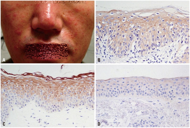
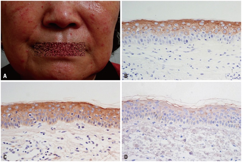
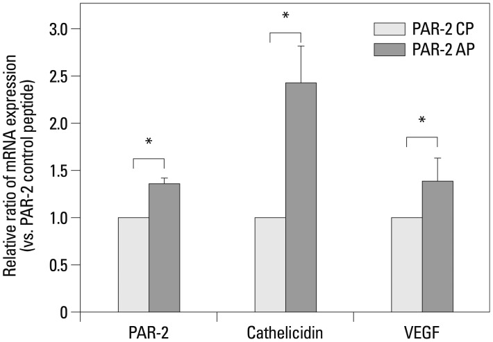
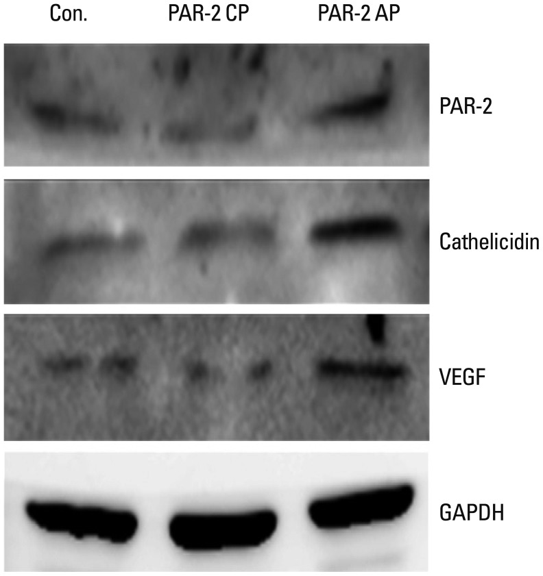
Materials and methods: Samples from 40 patients with clinicopathologic diagnosis of rosacea and facial skin tissue samples from 20 patients with no specific findings or milium without inflammation were retrieved. Intensities of immunohistochemical staining for PAR-2 and cathelicidin were compared between normal and rosacea-affected skin tissues. Additionally, correlations between PAR-2 and cathelicidin staining intensities within rosacea patients were analyzed. In cultured keratinocytes, changes in PAR-2, cathelicidin, and vascular endothelial growth factor (VEGF) mRNA and protein were analyzed after treatment with PAR-2 activating peptide (AP).
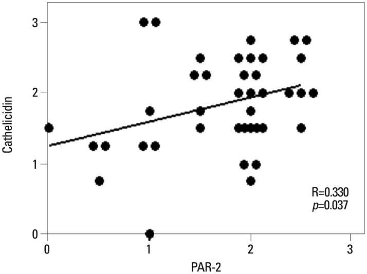
Results: Cathelicidin expression was significantly higher in rosacea skin tissues than in normal tissues (p<0.001), while PAR-2 expression was not significantly higher in rosacea tissues than in normal skin tissues. A positive correlation between PAR-2 and cathelicidin within rosacea samples was observed (R=0.330, p=0.037). After treatment of PAR-2 AP, both mRNA and protein levels for PAR-2, cathelicidin, and VEGF significantly increased in cultured keratinocytes, compared with PAR-2 control peptide treatment.
Conclusion: PAR-2 may participate in the pathogenesis of rosacea through activation of cathelicidin LL-37, a mediator of innate immune responses in the skin.
Keywords: Cathelicidin; LL-37; PAR-2; VEGF; rosacea.
Conflict of interest statement
The authors have no financial conflicts of interest.
Figures
Similar articles
-
Light-emitting diodes downregulate cathelicidin, kallikrein and toll-like receptor 2 expressions in keratinocytes and rosacea-like mouse skin.Exp Dermatol. 2016 Dec;25(12):956-961. doi: 10.1111/exd.13133. Epub 2016 Oct 3. Exp Dermatol. 2016. PMID: 27315464
-
Increased serine protease activity and cathelicidin promotes skin inflammation in rosacea.Nat Med. 2007 Aug;13(8):975-80. doi: 10.1038/nm1616. Epub 2007 Aug 5. Nat Med. 2007. PMID: 17676051
-
Coptis chinensis Franch Directly Inhibits Proteolytic Activation of Kallikrein 5 and Cathelicidin Associated with Rosacea in Epidermal Keratinocytes.Molecules. 2020 Nov 26;25(23):5556. doi: 10.3390/molecules25235556. Molecules. 2020. PMID: 33256158 Free PMC article.
-
[New insights in the pathogenesis and treatment of rosacea].Duodecim. 2012;128(22):2327-35. Duodecim. 2012. PMID: 23342479 Review. Finnish.
-
[Cathelicidin LL-37. A central factor in the pathogenesis of inflammatory dermatoses?].Hautarzt. 2008 Jan;59(1):72-4. doi: 10.1007/s00105-007-1457-z. Hautarzt. 2008. PMID: 18209993 Review. German.
Cited by
-
Genetic analyses reveal association between rosacea and cardiovascular diseases.Sci Rep. 2025 Feb 24;15(1):6657. doi: 10.1038/s41598-025-91240-4. Sci Rep. 2025. PMID: 39994418 Free PMC article.
-
Current research and clinical trends in rosacea pathogenesis.Heliyon. 2022 Oct 13;8(10):e10874. doi: 10.1016/j.heliyon.2022.e10874. eCollection 2022 Oct. Heliyon. 2022. PMID: 36276718 Free PMC article.
-
MMP-9 Levels in the Gingival Crevicular Fluid of Chilean Rosacea Patients.Int J Mol Sci. 2022 Aug 30;23(17):9858. doi: 10.3390/ijms23179858. Int J Mol Sci. 2022. PMID: 36077255 Free PMC article.
-
Consensus on the therapeutic management of rosacea - Brazilian Society of Dermatology.An Bras Dermatol. 2020 Nov-Dec;95 Suppl 1(Suppl 1):53-69. doi: 10.1016/j.abd.2020.08.001. Epub 2020 Oct 10. An Bras Dermatol. 2020. PMID: 33172727 Free PMC article.
-
Rosacea: Molecular Mechanisms and Management of a Chronic Cutaneous Inflammatory Condition.Int J Mol Sci. 2016 Sep 15;17(9):1562. doi: 10.3390/ijms17091562. Int J Mol Sci. 2016. PMID: 27649161 Free PMC article. Review.
References
-
- Korting HC, Schöllmann C. Current topical and systemic approaches to treatment of rosacea. J Eur Acad Dermatol Venereol. 2009;23:876–882. - PubMed
-
- Wilkin J, Dahl M, Detmar M, Drake L, Feinstein A, Odom R, et al. Standard classification of rosacea: Report of the National Rosacea Society Expert Committee on the Classification and Staging of Rosacea. J Am Acad Dermatol. 2002;46:584–587. - PubMed
-
- Yamasaki K, Gallo RL. Rosacea as a disease of cathelicidins and skin innate immunity. J Investig Dermatol Symp Proc. 2011;15:12–15. - PubMed
-
- Nizet V, Ohtake T, Lauth X, Trowbridge J, Rudisill J, Dorschner RA, et al. Innate antimicrobial peptide protects the skin from invasive bacterial infection. Nature. 2001;414:454–457. - PubMed
-
- Zanetti M. The role of cathelicidins in the innate host defenses of mammals. Curr Issues Mol Biol. 2005;7:179–196. - PubMed
Publication types
MeSH terms
Substances
LinkOut - more resources
Full Text Sources
Other Literature Sources
Medical
