Review
. 2015 Jan;20(1):1-11.
doi: 10.1007/s00776-014-0653-9. Epub 2014 Oct 17.

Clinical pathways for fragility fractures of the pelvic ring: personal experience and review of the literature

Affiliations
Review

Clinical pathways for fragility fractures of the pelvic ring: personal experience and review of the literature

Pol M Rommens et al. J Orthop Sci. 2015 Jan.

Abstract

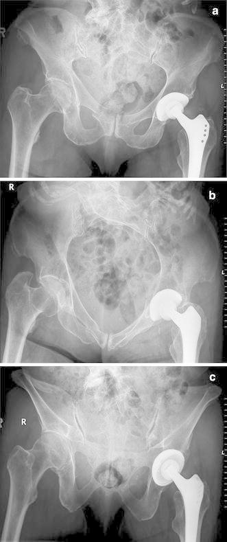
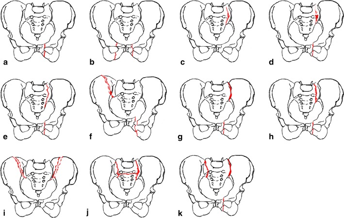
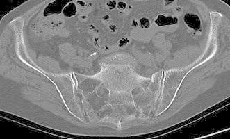
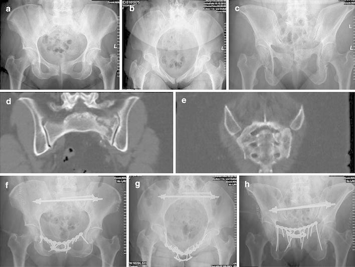
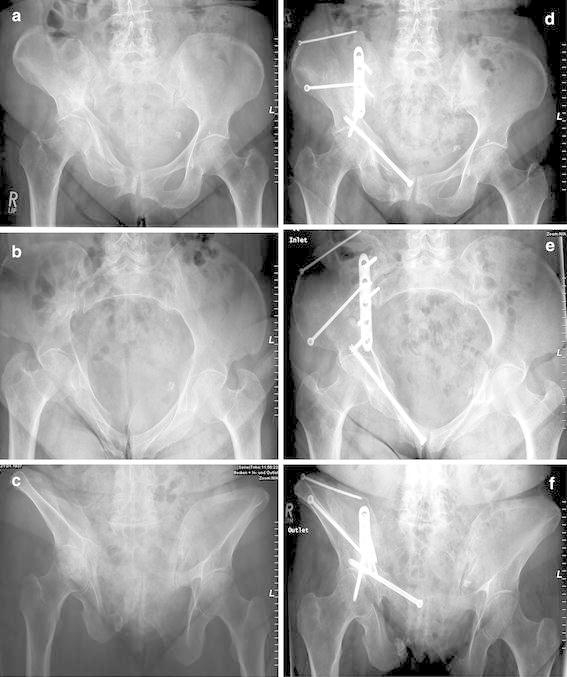
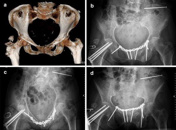
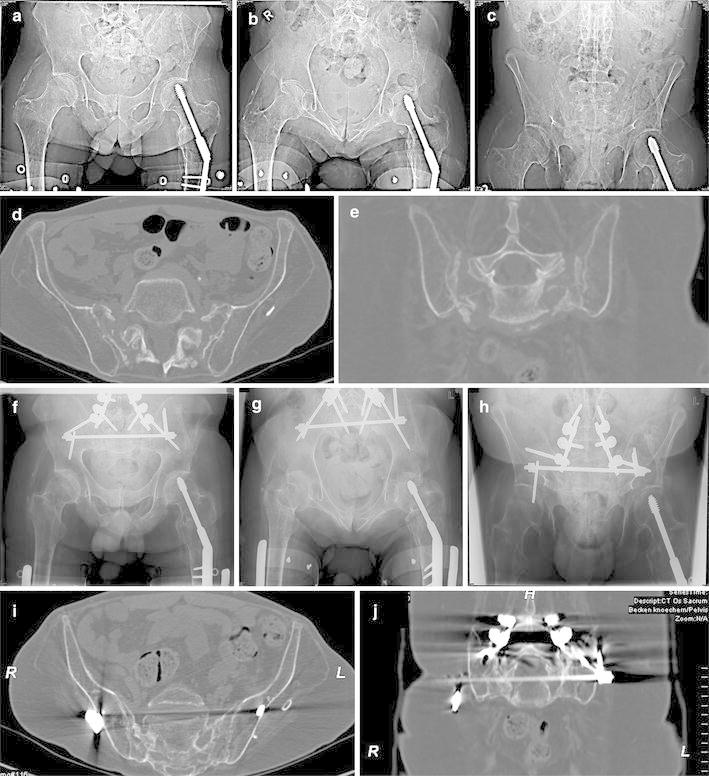
Fragility fractures of the pelvic ring (FFP) are increasing in frequency and require challenging treatment. A new comprehensive classification considers both fracture morphology and degree of instability. The classification system also provides recommendations for type and invasiveness of treatment. In this article, a literature review of treatment alternatives is presented and compared with our own experiences. Whereas FFP Type I lesions can be treated conservatively, FFP Types III and IV require surgical treatment. For FFP Type II lessions, percutaneous fixation techniques should be considered after a trial of conservative treatment. FFP Type III lesions need open reduction and internal fixation, whereas FFP Type IV lesions require bilateral fixation. The respective advantages and limitations of dorsal (sacroiliac screw fixation, sacroplasty, bridging plate fixation, transsacral positioning bar placement, angular stable plate) and anterior (external fixation, angular stable plate fixation, retrograde transpubic screw fixation) pelvic fixations are described.


Figures

Fig. 1
Fig. 1
a Pelvic ap overview of Type Ia FFP in an 82-year old woman. There is slight overriding of the fracture fragments at the right superior pubic ramus; b Pelvic inlet overview. A slight internal rotation of the right hemipelvis at the fracture of the right anterior pelvic ring is visible; c Pelvic outlet overview. The fracture of the right anterior pubic ramus is hardly visible. There is no lesion of the posterior pelvic ring
Fig. 2
Fig. 2
Classification of fragility fractures of the pelvis. a FFP Type Ia: unilateral anterior pelvic ring disruption. b FFP Type Ib: bilateral anterior pelvic ring disruption. c FFP Type IIa: dorsal non-displaced posterior injury only. d FFP Type IIb: sacral crush with anterior disruption. e FFP Type IIc: non-displaced sacral, sacroiliac or iliac fracture with anterior disruption. f FFP Type IIIa: displaced unilateral ilium fracture and anterior disruption. g FFP Type IIIb: displaced unilateral sacroiliac disruption and anterior disruption. h FFP Type IIIc: displaced unilateral sacral fracture together with anterior disruption. i FFP Type IVa: bilateral iliac fractures or bilateral sacroiliac disruptions together with anterior disruption. j FFP Type IVb: spinopelvic dissociation with anterior disruption. k FFP Type IVc: combination of different posterior instabilities together with anterior disruption
Fig. 3
Fig. 3
Transverse CT cut through dorsal pelvis of 83-year old female. Zones of low bone density in both sacral alae and a fracture of the left sacral ala are visible
Fig. 4
Fig. 4
a Pelvic ap overview of 72-year old female with anterior and posterior intervertebral fusion between L4 and S1. The patient had a history of more than three months of severe pain after a fall at home. A bilateral fracture of the pubic rami is visible with sclerotic margins, demonstrating a non-union. A fracture of the dorsal pelvic ring is not clear; b Axial CT reconstruction shows a bilateral fracture of the sacral ala at the S1 level; c Coronal reconstruction gives another view of the bilateral sacral ala lesions. This is an unstable lesion of the pelvic ring classified as FFP Type IVb; d Pelvic ap overview five months after operative reconstruction. Two sacroliliac screws have been inserted in the S1 body bilaterally. Insertion of a transsacral bar was not possible due to the pedicular screws in S1. The bilateral anterior instability was bridged with a long plate and screws construct; e Pelvic inlet overview; f Pelvic outlet overview. Note the long screws into the posterior column providing stability in this osteoporotic bone. The patient is free of complaints in the pelvic region and able to walk without crutches
Fig. 5
Fig. 5
Seventy-two-year old female with an FFP IIc lesion. a Pelvic ap overview. There are irregularities and a slight displacement at the symphysis pubis. A clear lesion of the dorsal pelvis is not visible; b Pelvic inlet view; c Pelvic outlet view. Instability of the symphysis pubis is visible as a step-off; d Coronal CT reconstruction of the dorsal pelvis. There is an undisplaced, yet complete, fracture of the left massa lateralis of the sacrum; e The fracture of the left lateral mass of the sacrum runs down through the neuroforamina S1 and S2; f Postoperative pelvic ap view. A sacral bar has been placed through the body of S1. On the left side, an additional sacroiliac screw has been placed. The instability of the symphysis pubis has been fixed with a double plate osteosynthesis and long screws into the posterior column, the anterior plate being angular stable; g Postoperative pelvic inlet view; h Postoperative pelvic outlet view. Three months after surgery, the patient was walking independently without complaints at the pelvis
Fig. 6
Fig. 6
a Seventy-four-year old female with an FFP Type IIIa lesion. A complete right-sided crescent fracture of the ilium and superior and inferior pubic ramus fractures with vertical displacement are visible in the pelvic a.p. view; b Pelvic inlet view; c Pelvic outlet view; d Postoperative pelvic a.p. view. The ilium fracture has been fixed with a large fragment angular stable plate and two long lag screws. The pubic ramus fracture has been splinted with a minimally invasive retrograde transpubic screw; e Postoperative pelvic inlet view; f Postoperative pelvic outlet view
Fig. 7
Fig. 7
a Surface rendering of the pelvic bone in an 87-year old female after a fall at home. An incomplete fracture of the dorsal ilium, a displaced fracture of the superior pubis ramus and an undisplaced inferior pubic ramus fracture are visible. The very low bone density in both sacral ala and in the center of the iliac wing are clearly visible. The lesion is classified as an FFP Type IIc; b Postoperative pelvic a.p. overview. The incomplete ilium fracture has been fixed with a single screw, the anterior instability with a bridging plate and screw osteosynthesis. To avoid loosening of the screws, an we chose the longest possible screw trajectory in the pubic bone and ischium; c Postoperative pelvic inlet overview; d Postoperative pelvic outlet overview. Six months after surgery, the patient walked independently and without complaints
Fig. 8
Fig. 8
a Pelvic a.p. overview of a 55-year old patient who became paraplegic at the age of 27. There is a rarefication of bone substance in the pelvic bone and lower extremities. The dorsal pelvis is difficult to appreciate; b Pelvic inlet overview. The sacrum seems intruded into the pelvic ring; c Pelvic outlet overview. The lumbosacral segment seems displaced distally when related to the pelvic ring; d Axial CT-reconstruction of the dorsal pelvis showing a severe rarefication of the bone substance and a complete bilateral fragility fracture of the lateral mass of the sacrum; e Coronal CT-reconstruction confirms the complete bilateral fragility fractures of the lateral mass of the sacrum; f Postoperative pelvic a.p. overview. An iliolumbar fusion between L4, L5 and the dorsal ilium was done. Additionally, a transsacral positioning bar was placed through the body of S1. Small plates were used as washers under the nuts of the sacral bar to hinder cut-through of the washers; g Postoperative pelvic inlet view; h Postoperative pelvic outlet view; i A representative axial CT-cut one year after surgery, showing complete healing of the sacral fragility fractures; j A representative coronal CT-cut showing the position of the transpedicular screws and the transsacral positioning bar. The healing of the sacrum is visible. The patient is able to stand with auxiliary means

References

    1. World factbook: https://www.cia.gov/library/publications/the-world-factbook/rankorder/21....
    1. Dhanwal DK, Dennison EM, Harvey NC, Cooper C. Epidemiology of hip fracture: worldwide geographic variation. Indian J Orthop. 2011;45(1):15–22. doi: 10.4103/0019-5413.73656. - DOI - PMC - PubMed
    1. Watts NB. The Fracture Risk Assessment Tool (FRAX®): applications in clinical practice. J Womens Health (Larchmt) 2011;20(4):525–531. doi: 10.1089/jwh.2010.2294. - DOI - PubMed
    1. Johnell O, Kanis JA. An estimate of the worldwide prevalence and disability associated with osteoporotic fractures. Osteoporos Int. 2006;17(12):1726–1733. doi: 10.1007/s00198-006-0172-4. - DOI - PubMed
    1. Kates SL, Bukata SV, DiGiovanni BF, Friedman SM, Hoyen H, Kates A, Kates SL, Mears SC, Mendelson DA, Serna FH, Jr., Sieber FE and Tyler WK. A guide to improving the care of patients with fragility fractures. Geriatric orthopaedic surgery and rehabilitation 2011; 2: 5-37. http://gos.sagepub.com/content/2/1/5. - PMC - PubMed