Correlation between human tear cytokine levels and cellular corneal changes in patients with bacterial keratitis by in vivo confocal microscopy
- PMID: 25324281
- PMCID: PMC4240721
- DOI: 10.1167/iovs.14-15411
Correlation between human tear cytokine levels and cellular corneal changes in patients with bacterial keratitis by in vivo confocal microscopy
Abstract
Purpose: We investigated bilateral tear cytokine levels in patients with unilateral bacterial keratitis (BK) as associated with in vivo confocal microscopic (IVCM) alterations in corneal nerves and dendritiform immune cells (DCs).
Methods: A total of 54 (13 BK, 13 contralateral, 28 healthy controls) tear samples was collected prospectively and analyzed by multiplex microbeads assay. The IVCM of the central cornea was performed on the same day, and assessed for corneal nerve and DC alterations.
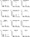
Results: Interleukin-1β, IL-6, and IL-8 were significantly elevated only in affected eyes (66.6 ± 26.8, 7174 ± 2430, and 810 ± 315 ρg/mL, respectively; P = 0.04, P < 0.001, and P < 0.001, respectively), compared to healthy controls (13.0 ± 4.0, 171.8 ± 32.1, and 56.5 ± 33.8 ρg/mL). Levels of chemokine ligand 2 (CCL-2), IL-10, and IL-17a were elevated only in contralateral eyes (813 ± 478, 86.7 ± 38.3, and 3350 ± 881 ρg/mL, respectively; P = 0.02, P = 0.01, and P = 0.04, respectively), compared to controls (73.7 ± 25.3, 17.5 ± 4.9, and 1350 ± 337 ρg/mL). Triggering receptor expressed on myeloid cells (TREM)-1 was significantly elevated in affected (551 ± 231 ρg/mL, P = 0.02) and contralateral unaffected (545 ± 298 ρg/mL, P = 0.03) eyes compared to controls (31.3 ± 12.4 ρg/mL). The density of DCs was significantly increased in affected (226.9 ± 37.3 cells/mm(2), P < 0.001) and unaffected (122.3 ± 23.7 cells/mm(2), P < 0.001) eyes compared to controls (22.7 ± 5.9 cells/mm(2)). Sub-basal nerve density significantly decreased in affected (3337 ± 1615 μm/mm(2), P < 0.001) and contralateral (13,230 ± 1635 μm/mm(2), P < 0.001) eyes compared to controls (21,200 ± 545 μm/mm(2)). Levels of IL-1β, IL-6, and IL-8 were significantly correlated with DC density (R = 0.40, R = 0.55, and R = 0.31, all P < 0.02) and nerve density (R = -0.30, R = -0.53, and R = -0.39, all P < 0.01).
Conclusions: Proinflammatory tear cytokines are elevated bilaterally in patients with unilateral BK, and are correlated strongly with alterations in DCs and nerve density as detected by IVCM.
Keywords: corneal infection; cytokines; dendritic cells; infectious keratitis; tears.
Copyright 2014 The Association for Research in Vision and Ophthalmology, Inc.
Figures


Similar articles
-
Contralateral Clinically Unaffected Eyes of Patients With Unilateral Infectious Keratitis Demonstrate a Sympathetic Immune Response.Invest Ophthalmol Vis Sci. 2015 Oct;56(11):6612-20. doi: 10.1167/iovs.15-16560. Invest Ophthalmol Vis Sci. 2015. PMID: 26465889 Free PMC article.
-
Bilateral Alterations in Corneal Nerves, Dendritic Cells, and Tear Cytokine Levels in Ocular Surface Disease.Cornea. 2016 Nov;35 Suppl 1(Suppl 1):S65-S70. doi: 10.1097/ICO.0000000000000989. Cornea. 2016. PMID: 27617877 Free PMC article. Review.
-
Inflammation and the nervous system: the connection in the cornea in patients with infectious keratitis.Invest Ophthalmol Vis Sci. 2011 Jul 11;52(8):5136-43. doi: 10.1167/iovs.10-7048. Invest Ophthalmol Vis Sci. 2011. PMID: 21460259 Free PMC article.
-
In Vivo Confocal Microscopy Demonstrates Bilateral Loss of Endothelial Cells in Unilateral Herpes Simplex Keratitis.Invest Ophthalmol Vis Sci. 2015 Jul;56(8):4899-906. doi: 10.1167/iovs.15-16527. Invest Ophthalmol Vis Sci. 2015. PMID: 26225629 Free PMC article.
-
Applications of in vivo confocal microscopy in the management of infectious keratitis in veterinary ophthalmology.Vet Ophthalmol. 2022 May;25 Suppl 1:5-16. doi: 10.1111/vop.12928. Epub 2021 Sep 4. Vet Ophthalmol. 2022. PMID: 34480385 Review.
Cited by
-
Defining an Optimal Sample Size for Corneal Epithelial Immune Cell Analysis Using in vivo Confocal Microscopy Images.Front Med (Lausanne). 2022 Jun 1;9:848776. doi: 10.3389/fmed.2022.848776. eCollection 2022. Front Med (Lausanne). 2022. PMID: 35721066 Free PMC article.
-
Changes in corneal higher-order aberrations during treatment for infectious keratitis.Sci Rep. 2023 Jan 16;13(1):848. doi: 10.1038/s41598-023-28145-7. Sci Rep. 2023. PMID: 36646747 Free PMC article.
-
The Role of Connexin-43 in the Inflammatory Process: A New Potential Therapy to Influence Keratitis.J Ophthalmol. 2019 Jan 21;2019:9312827. doi: 10.1155/2019/9312827. eCollection 2019. J Ophthalmol. 2019. PMID: 30805212 Free PMC article. Review.
-
Expression of IL-8, IL-6 and IL-1β in tears as a main characteristic of the immune response in human microbial keratitis.Int J Mol Sci. 2015 Mar 3;16(3):4850-64. doi: 10.3390/ijms16034850. Int J Mol Sci. 2015. PMID: 25741769 Free PMC article.
-
The Innate Immune Cell Profile of the Cornea Predicts the Onset of Ocular Surface Inflammatory Disorders.J Clin Med. 2019 Dec 2;8(12):2110. doi: 10.3390/jcm8122110. J Clin Med. 2019. PMID: 31810226 Free PMC article.
References
-
- Prokosch V, Gatzioufas Z, Thanos S, Stupp T. Microbiological findings and predisposing risk factors in corneal ulcers. Graefes Arch Clin Exp Ophthalmol. 2008; 250: 369–374. - PubMed
-
- Green M, Apel A, Stapleton F. Risk factors and causative organisms in microbial keratitis. Cornea. 2008; 27: 22–27. - PubMed
-
- Webb RM, Duke MA. Bacterial infection of a neurotrophic cornea in an immunocompromised subject. Cornea. 1985; 4: 14–18. - PubMed
Publication types
MeSH terms
Substances
Grants and funding
LinkOut - more resources
Full Text Sources
Other Literature Sources
Medical

