. 2014 Nov 10;42(20):12483-97.
doi: 10.1093/nar/gku953. Epub 2014 Oct 16.

hnRNPA1 couples nuclear export and translation of specific mRNAs downstream of FGF-2/S6K2 signalling

Affiliations

hnRNPA1 couples nuclear export and translation of specific mRNAs downstream of FGF-2/S6K2 signalling

Rajat Roy et al. Nucleic Acids Res. .

Erratum in

Abstract

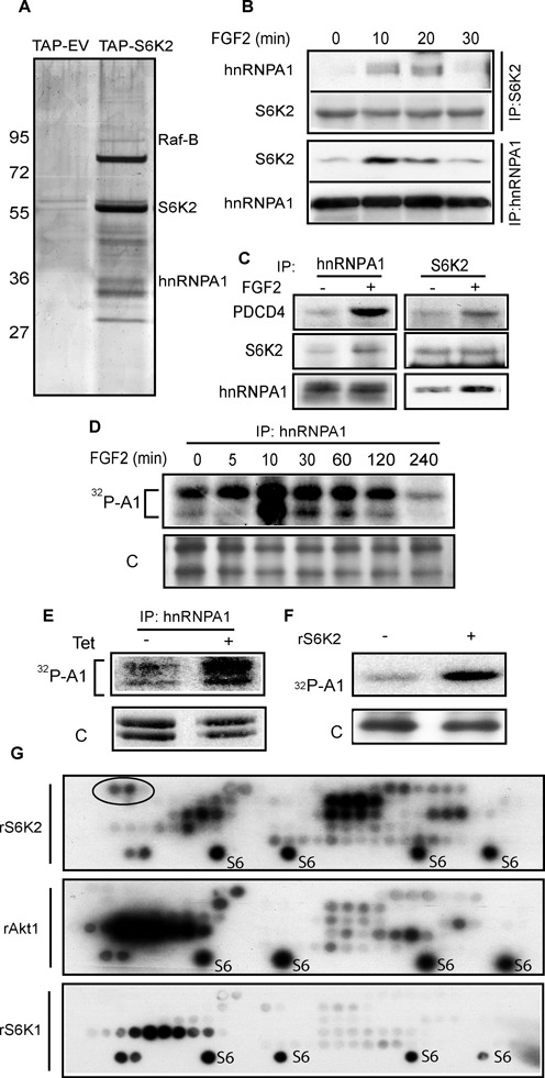
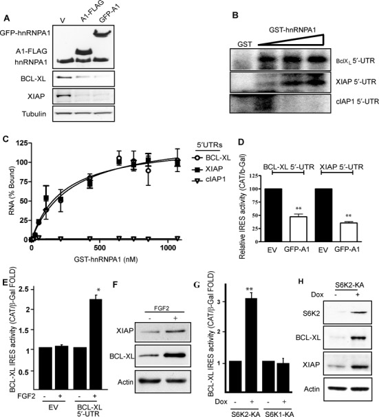
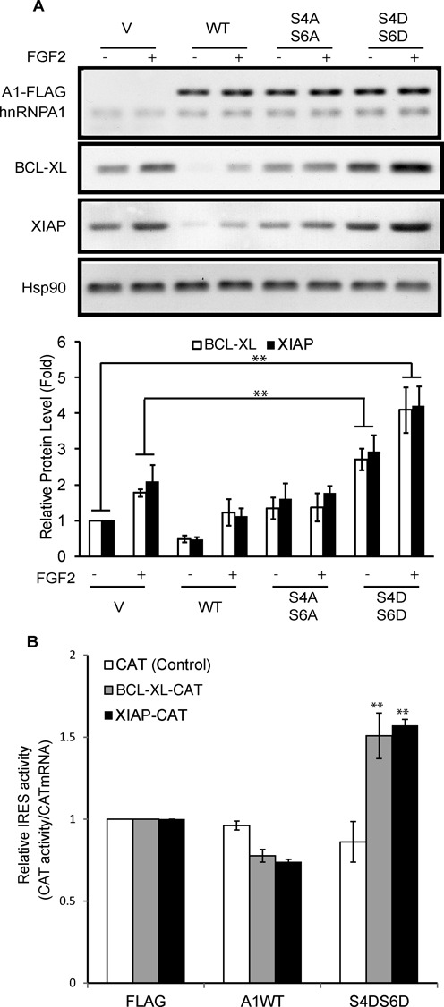
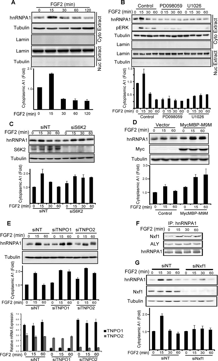
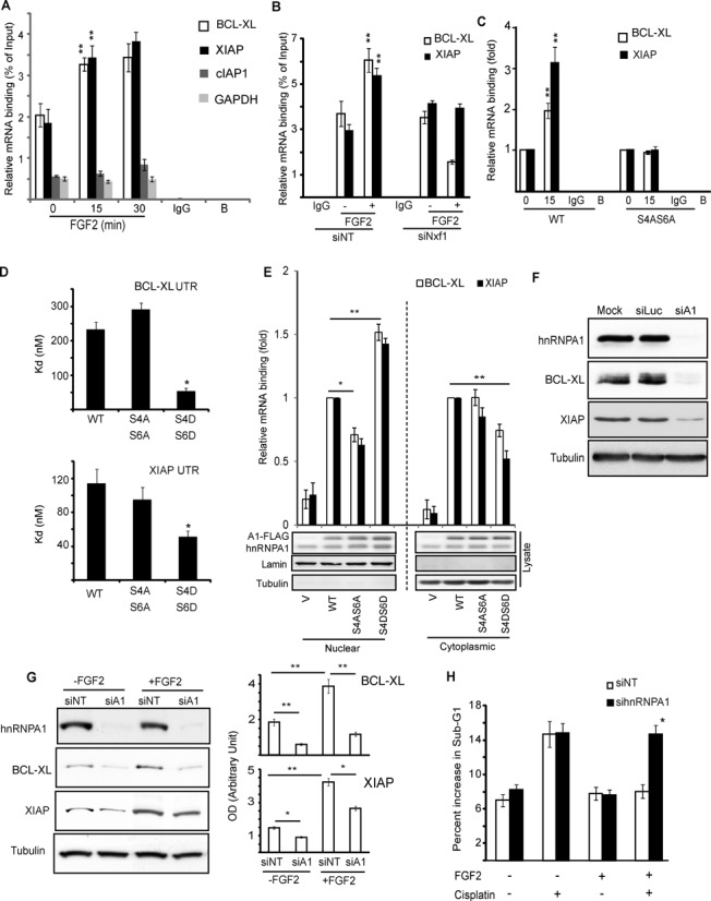
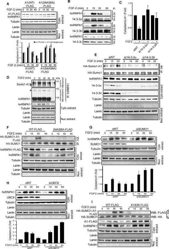
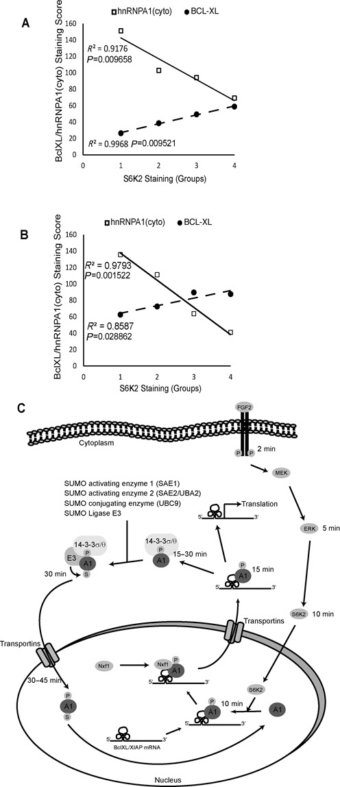
The increased cap-independent translation of anti-apoptotic proteins is involved in the development of drug resistance in lung cancer but signalling events regulating this are poorly understood. Fibroblast growth factor 2 (FGF-2) signalling-induced S6 kinase 2 (S6K2) activation is necessary, but the downstream mediator(s) coupling this kinase to the translational response is unknown. Here, we show that S6K2 binds and phosphorylates hnRNPA1 on novel Ser4/6 sites, increasing its association with BCL-XL and XIAP mRNAs to promote their nuclear export. In the cytoplasm, phosphoS4/6-hnRNPA1 dissociates from these mRNAs de-repressing their IRES-mediated translation. This correlates with the phosphorylation-dependent association of hnRNPA1 with 14-3-3 leading to hnRNPA1 sumoylation on K183 and its re-import into the nucleus. A non-phosphorylatible, S4/6A mutant prevented these processes, hindering the pro-survival activity of FGF-2/S6K2 signalling. Interestingly, immunohistochemical staining of lung and breast cancer tissue samples demonstrated that increased S6K2 expression correlates with decreased cytoplasmic hnRNPA1 and increased BCL-XL expression. In short, phosphorylation on novel N-term sites of hnRNPA1 promotes translation of anti-apoptotic proteins and is indispensable for the pro-survival effects of FGF-2.


Figures

Figure 1.
Figure 1.
hnRNPA1 interacts with and is phosphorylated by S6K2. (A) HEK293 cells expressing TAP-tagged S6K2 were subjected to Tandem Affinity Purification, proteins separated by SDS-PAGE and the silver stained bands identified by MS. (B and C) S6K2 or hnRNPA1 were immunoprecipitated from H510 (B) or HEK293 (C) cells treated with FGF-2 and associated proteins detected by western blot. (D) HEK293 cells were labelled with 32Pi phosphate prior to treatment with FGF-2 and hnRNPA1 immunoprecipitation. The samples were separated by SDS-PAGE and autoradiographed. (E) HEK293 cells expressing tetracycline-inducible kinase-active S6K2 were labelled with 32Pi phosphate and hnRNPA1 immunoprecipitation performed post treatment ± doxycline (Dox). The samples were separated on SDS-PAGE prior to autoradiography. (F) Recombinant (r) hnRNPA1 and S6K2 were used in an in vitro kinase (IVK) assay in presence of 32P γATP, prior to SDS-PAGE and autoradiography. (D, E and F) ‘C’; silver stain control. (G) hnRNPA1 peptide arrays were spotted on nitrocellulose membrane and IVK assays performed with rS6K2, rS6K1 or rAkt1 in presence of 32P γATP prior to autoradiography. Ribosomal S6 peptide (S6) was used as a positive control. Two peptides uniquely phosphorylated by S6K2 are circled. All experiments are representative of at least three independent repeats. See also Supplementary Tables S1–S4 and Supplementary Figure S1.
Figure 2.
Figure 2.
hnRNPA1 binds to BCL-XL, XIAP transcripts in vitro and regulates their translation. (A) N-term GFP- and C-term FLAG-tagged hnRNPA1 were transiently expressed in HEK293 cells and western blotting (WB) performed for the indicated proteins 72 h later. (B and C) Recombinant GST-hnRNP A1 was incubated in the presence of 32P-labelled, in vitro transcribed BCL-XL, XIAP or cIAP1 (negative control) 5′-UTR RNAs and UV crosslinked. (B) RNA–protein complexes were analysed by SDS-PAGE/autoradiography. GST was used as a negative control. (C) Increasing concentrations of GST-hnRNPA1 were used to bind the UTRs and the signal from nitrocellulose filter binding assays quantified by beta counter. (D, E and G) Bicistronic DNA constructs containing XIAP or BCL-XL IRES elements were co-transfected into (D) HEK293 cells along with GFP (EV) or GFP-hnRNPA1 (GFP-A1) expressing plasmids, (E) transfected into HEK293 cells treated ± FGF-2 for 4 h or (G) into HEK293 cells stably expressing tetracycline-inducible kinase-active (KA) S6K1 or 2 treated overnight ± doxycline (Dox). IRES activity was measured as ratio of CAT to β-gal expression. (F and H) HEK293 cells (F) or S6K2-KA HEK293 cells (H) were stimulated ± FGF-2 for 4 h or overnight ± Dox, respectively, and samples analysed by WB for the indicated proteins. (C, D, E and G) Results are the average of triplicates ± SEM. Student's t-test: *P < 0.05; **P < 0.01.
Figure 3.
Figure 3.
Phosphorylation of Ser4/6 on hnRNPA1 regulates BCL-XL and XIAP expression. (A and B) HEK293 cells were transiently transfected with wild-type (WT), mutant (S4AS6A or S4DS6D) hnRNPA1 or vector alone (V). (A) Cells were treated ± FGF-2 for 4 h prior to SDS-PAGE/western blotting for the indicated proteins. Lower panel: the BCL-XL and XIAP signals were quantified and normalized to that of HSP90. (B) Cells were subsequently transfected with reporter mRNAs driving CAT from the BCL-XL or XIAP IRES. CAT activity was normalised to the amount of injected mRNA. CAT alone mRNAs were used as control. Results are average of (A, lower panel) three independent experiments ± SEM or (B) triplicates ± SEM. Student's t-test: *P < 0.05; **P < 0.01.
Figure 4.
Figure 4.
hnRNPA1 shuttles between cytoplasm and nucleus in response to FGF-2. HEK293 cells alone (A), pre-treated with the MEK inhibitors PD098059, U1026 or DMSO alone for 1 h (B), transfected with siRNA for S6K2 (C), TNPO1 and 2 (E) or NXF1 (G) or the karyopherin inhibitor MycMBP-M9M vector or vector alone (D) were treated ± FGF-2 for the indicated time. The cytoplasmic fraction was analysed by SDS-PAGE/western blotting (WB) for the indicated proteins. S6K2 (C), TNPO1 and 2 (E) or NXF1 (G) knockdown were confirmed by SDS-PAGE/WB (C and G) or qPCR (E, lower panel). (A–E and G Lower panel) The hnRNPA1 signal was quantified and normalized to that of tubulin. (F) Endogenous hnRNPA1 was immunoprecipitated from HEK293 cells treated ± FGF-2. Immunoprecipitates were analysed by SDS-PAGE/WB for the indicated proteins. (A–G) Results are representative of at least three independent experiments. Bar graphs are average from replicate experiments ± SEM. See also Supplementary Figure S2.
Figure 5.
Figure 5.
hnRNPA1 mediates nuclear export of BCL-XL and XIAP mRNA in response to FGF-2. (A) HEK293 cells, (B) HEK293 cells transfected with Nxf1 siRNA or non-targeting control (NT) siRNA or (C) HEK293 cells expressing wild-type or S4AS6A mutant hnRNPA1 were treated with FGF-2 for the indicated durations (A and C) or 30 min (B). The cytoplasmic fractions were subjected to RNA immunoprecipitation (RNA-IP) with hnRNPA1 antibody and the associated RNA analysed by quantitative PCR (qPCR) using primers directed against the indicated transcripts. (D) Increasing concentrations of recombinant wild-type (WT), S4AS6A or S4DS6D mutant hnRNPA1 were incubated with 32P-labelled, in vitro transcribed XIAP or BCL-XL 5′-UTR, nitrocellulose filter binding assays performed and binding quantified as in Figure 2C. Kd values were calculated from three independent experiments using non-linear one-site specific binding. (E) HEK293 cells were transiently transfected with WT, S4AS6A, S4DS6D hnRNPA1 or vector alone (V) and subcellular fractionation was performed 48 h later to separate the nuclear and cytoplasmic fractions. These were subjected to RNA-IP and qPCR as in (A). Results are expressed as average fold changes from triplicate ± SEM with cells expressing WT-hnRNPA1 as reference. (F) HEK293 cells were transfected with hnRNPA1 (siA1) or luciferase (siLuc) control siRNAs or mock transfected (transfection reagent alone). Cell lysates were analysed by SDS-PAGE/western blotting (WB). (G) HEK293 cells transfected ± hnRNPA1 or non-targeting (siNT) siRNAs and treated ± FGF-2 for 4 h followed or not by treatment with cisplatin for 48 h were (G) analysed by SDS-PAGE/WB for their BCL-XL and XIAP levels or (H) analysed by flow cytometry following PI staining for the appearance of a Sub-G1 (apoptotic) population. Results are average of triplicates ± SEM. (G) Left panel: average of intensities for BCL-XL and XIAP bands from three experiments were normalized to that of tubulin and plotted ± SEM. (A–H) Results representative of at least three independent experiments. Student's t-test: *P < 0.05; **P < 0.01.
Figure 6.
Figure 6.
Cytoplasmic hnRNPA1 associates with 14-3-3 subunits and is sumoylated upon stimulation with FGF-2. (A, C, G and H) HEK293 cells were transiently transfected with (A) wild-type (WT) or S4AS6A mutant hnRNPA1-FLAG or (C, G and H) siRNAs against the indicated proteins or a non-targeting control (NT) were treated ± FGF-2 for the indicated time (A and G) or 60 min (C) and the cytoplasmic fraction analysed by SDS-PAGE/western blotting (WB). (A, G and H lower panels and C) Band intensities for hnRNPA1 were quantified and normalized to that for tubulin. Results show average of triplicate experiments ± SEM normalised to the corresponding controls. In Fig6A, lower panel shows quantification for both endogenous and the FLAG-tagged hnRNPA1. (B) HEK293 cells were treated with FGF-2 and the endogenous 14-3-3 protein subunits σ, θ or ϵ immunoprecipitated (IP) from the cytoplasmic fraction. Immunoprecipitates were analysed by SDS-PAGE/WB for endogenous hnRNPA1 or 14-3-3 proteins. (D) Endogenous hnRNPA1 was immunoprecipitated from the cytoplasmic fraction of HEK293 cells treated ± FGF-2. Immunoprecipitates were analysed by SDS-PAGE/WB with either Sumo1 or hnRNPA1 antibodies. The arrows indicate sumoylated-hnRNPA1. (E) HEK293 cells stably expressing HA-Sumo1 were treated with siRNA for 14-3-3σ, θ or NT before stimulation with FGF-2. HA-SUMO1 was immunoprecipitated with anti-HA antibodies from the cytoplasmic fraction and the samples analysed by SDS-PAGE/WB for the indicated proteins. (F and I) HEK293 cells stably expressing HA-SUMO1 were transiently transfected with FLAG-tagged WT or S4AS6A or K183R mutant hnRNPA1 and treated with FGF-2. Cytoplasmic fractions were immunoprecipitated for HA-SUMO1 and analysed as above. All experiments are representative of three independent experiments. See also Supplementary Figure S3.
Figure 7.
Figure 7.
Immunohistochemical staining for S6K2, hnRNPA1 and BCL-XL. TMA samples from lung (n = 204) (A) and breast cancer (n = 194) (B) patients were stained for S6K2, hnRNPA1 and BCL-XL. An increase in S6K2 staining correlates with concomitant increase in BCL-XL and decrease in cytoplasmic hnRNPA1 staining. Patients were grouped (X axis; group 1–4) according to their staining scores for S6K2 and correlation curves between the staining intensities for BCL-XL and cytoplasmic hnRNPA1 with groups 1–4 plotted. Correlation coefficients and P-values (ANOVA test) are shown. (C) Schematic illustration of how FGF-2 signalling regulates the shuttling of hnRNPA1 and the translation of BCL-XL and XIAP mRNAs.

Similar articles

Cited by

References

    1. Li X.H., Li C., Xiao Z.Q. Proteomics for identifying mechanisms and biomarkers of drug resistance in cancer. J. Proteomics. 2011;74:2642–2649. - PubMed
    1. Indran I.R., Tufo G., Pervaiz S., Brenner C. Recent advances in apoptosis, mitochondria and drug resistance in cancer cells. Biochim. Biophys. Acta. 2011;1807:735–745. - PubMed
    1. Alama A., Orengo A.M., Ferrini S., Gangemi R. Targeting cancer-initiating cell drug-resistance: a roadmap to a new-generation of cancer therapies. Drug Discov. Today. 2012;17:435–442. - PubMed
    1. Pardo O.E., Wellbrock C., Khanzada U.K., Aubert M., Arozarena I., Davidson S., Bowen F., Parker P.J., Filonenko V.V., Gout I.T., et al. FGF-2 protects small cell lung cancer cells from apoptosis through a complex involving PKCepsilon, B-Raf and S6K2. EMBO J. 2006;25:3078–3088. - PMC - PubMed
    1. Pardo O.E., Arcaro A., Salerno G., Raguz S., Downward J., Seckl M.J. Fibroblast growth factor-2 induces translational regulation of Bcl-XL and Bcl-2 via a MEK-dependent pathway: correlation with resistance to etoposide-induced apoptosis. J. Biol. Chem. 2002;277:12040–12046. - PubMed

Publication types

MeSH terms

Substances