. 2014 Oct 17;346(6207):355-9.
doi: 10.1126/science.1259723. Epub 2014 Sep 25.

Structure and selectivity in bestrophin ion channels

Affiliations

Structure and selectivity in bestrophin ion channels

Tingting Yang et al. Science. .

Abstract

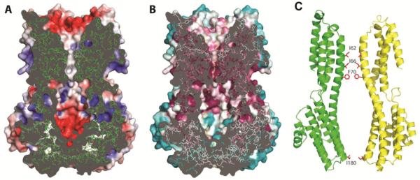
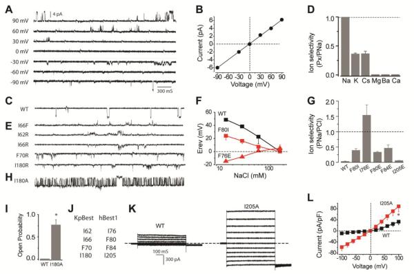
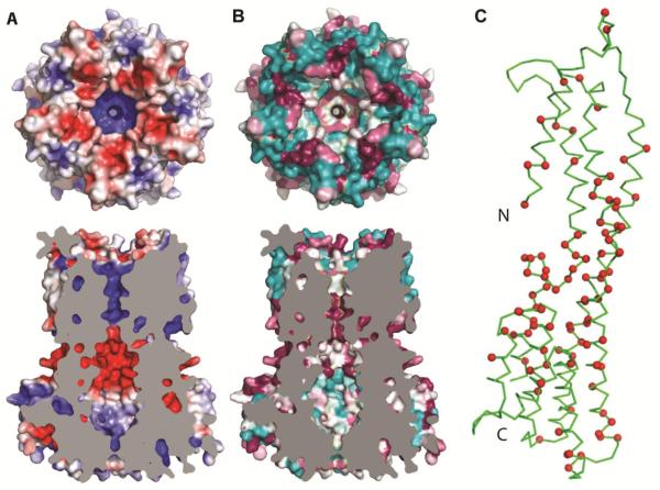
Human bestrophin-1 (hBest1) is a calcium-activated chloride channel from the retinal pigment epithelium, where mutations are associated with vitelliform macular degeneration, or Best disease. We describe the structure of a bacterial homolog (KpBest) of hBest1 and functional characterizations of both channels. KpBest is a pentamer that forms a five-helix transmembrane pore, closed by three rings of conserved hydrophobic residues, and has a cytoplasmic cavern with a restricted exit. From electrophysiological analysis of structure-inspired mutations in KpBest and hBest1, we find a sensitive control of ion selectivity in the bestrophins, including reversal of anion/cation selectivity, and dramatic activation by mutations at the cytoplasmic exit. A homology model of hBest1 shows the locations of disease-causing mutations and suggests possible roles in regulation.


Figures

Fig. 1
Fig. 1. Crystal structure of KpBest
(A & B) Ribbon diagram of the KpBest pentamer with each protomer colored differently, (A) as viewed from outside the membrane and (B) as viewed from the side (rotated 90° through × axis). (C & D) Electrostatic potential at the molecular surface viewed as in A & B, respectively). The contour level is at ±5 kT/e red for negative potential and blue for positive potential. Membrane boundaries in B & D were calculated by OPM server. (E) 2D topology of a protomer, colored spectrally from dark blue at its N-terminal segment to red at its C-terminal segment. (F) Ribbon diagram of a protomer. Colored as in E.
Fig. 2
Fig. 2. Structure of the ion conducting pathway through KpBest
(A) Cross-section through the pore center. The model is viewed as in 1D, with the electrostatic potential shown on exposed surfaces of the molecular envelope. (B) Cross-section as in A, but colored by Consurf sequence conservation. The calculation used 150 prokaryotic homologs with 95% maximal and 35% minimal sequence identities compared to KpBest. . (C) Ribbon diagram of two oppositely facing (144°) protomers of a KpBest pentamer are shown with the extracellular side on the top. The side chains of critical residues are red.
Fig. 3
Fig. 3. Ionic conductance measurements of KpBest and hBest1
(A) Representative families of single KpBest currents recorded from planar lipid bilayers at different voltages (150 mM NaCl in both trans and cis solutions). (B) Single KpBest channel current-voltage relationship. (C) Current trace of wild-type single KpBest channels (150 mM NaCl in cis and 0 NaCl in trans solutions). (D) Relative cation permeability; n = 3 for each point. (E) Current traces of mutant KpBest channels (same condition as C). (F) Changes in wild-type and mutant hBest1 channel reversal potentials (Erev) as a function of extracellular NaCl concentration (150 mM NaCl in internal); n=3−4 for each point. (G) Relative permeability (PNa/PCl) of hBest1 wild-type and mutant variants; n=12−15 for each bar. (H) Current trace of I180A single KpBest channels (same condition as C). (I) Wild-type and I180A single KpBest channel open probabilities. (J) Critical residues in KpBest and hBest1. (K) Exemplar whole-cell currents of wild type (left) and I205A (right) hBest1 in HEK 293 cells. (L) Population steady-state current-voltage relationships; n= 3−6 for each point. * P<0.05 when compared to wild-type using two-tailed unpaired Student's t test.
Fig. 4
Fig. 4. Homology model of hBest1
(A) Top, electrostatic potential at the extracellular surface of the hBest1 homology model. Viewed and drawn as for 1C. Bottom, cross-section through the homology model of hBest1. Viewed and drawn as for 2A, except that cut surface is plain grey. (B) Top, top view as in A, but colored by surface conservation. This calculation used 150 homologs with 95% maximal and 35% minimal sequence identities compared to hBest1. Bottom, cross-section viewed and drawn as in 2B but having a plain grey cut surface. (C) Ribbon diagram of a protomer from the hBest1 homology pentamer. The Cα positions of residues that are sites of disease-causing point mutations are marked in red.

References

    1. Marmorstein AD, Marmorstein LY, Rayborn M, Wang X, Hollyfield JG, Petrukhin K. Bestrophin, the product of the Best vitelliform macular dystrophy gene (VMD2), localizes to the basolateral plasma membrane of the retinal pigment epithelium. Proc Natl Acad Sci U S A. 2000;97:12758–12763. - PMC - PubMed
    1. Marquardt A, Stohr H, Passmore LA, Kramer F, Rivera A, Weber BH. Mutations in a novel gene, VMD2, encoding a protein of unknown properties cause juvenile-onset vitelliform macular dystrophy (Best's disease) Hum Mol Genet. 1998;7:1517–1525. - PubMed
    1. Petrukhin K, J. Koisti M, Bakall B, Li W, Xie G, Marknell T, Sandgren O, Forsman K, Holmgren G, Andreasson S, Vujic M, Bergen AA, McGarty-Dugan V, Figueroa D, Austin CP, Metzker ML, Caskey CT, Wadelius C. Identification of the gene responsible for Best macular dystrophy. Nat Genet. 1998;19:241–247. - PubMed
    1. Neussert R, Muller C, Milenkovic VM, Strauss O. The presence of bestrophin-1 modulates the Ca2+ recruitment from Ca2+ stores in the ER. Pflugers Archiv : European journal of physiology. 2010;460:163–175. - PubMed
    1. Boon CJ, Klevering BJ, Leroy BP, Hoyng CB, Keunen JE, den Hollander AI. The spectrum of ocular phenotypes caused by mutations in the BEST1 gene. Progress in retinal and eye research. 2009;28:187–205. - PubMed

Publication types