

Review
doi: 10.3174/ajnr.A4118.
Epub 2014 Oct 16.
Spinal cord ischemia: practical imaging tips, pearls, and pitfalls
Affiliations
- PMID: 25324492
- PMCID: PMC7990611
- DOI: 10.3174/ajnr.A4118
Item in Clipboard
Review
Spinal cord ischemia: practical imaging tips, pearls, and pitfalls
AJNR Am J Neuroradiol.
2015 May.
Abstract
Ischemia of the spinal cord is a rare entity with a poor prognosis. Brain ischemia is no longer a diagnostic challenge; on the contrary, ischemia of the spinal cord remains difficult, particularly in children. In this article, we illustrate the principal causes in children and adults, clinical presentation, different techniques for the diagnosis by MR imaging (diffusion, spinal MR angiography, and 1.5 versus 3T), pathophysiology, and differential diagnosis. We will discuss current knowledge, perspectives, and pitfalls.
© 2015 by American Journal of Neuroradiology.
Figures

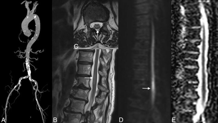
Localization of the artery of Adamkiewicz in a patient with aortic thrombus. MR angiography shows the thrombus in the abdominal aorta below the renal arteries (arrows, A). No ischemia is visible in the conus medullaris (B). The artery of Adamkiewicz is permeable (arrows, C).
Ischemia provoked by an atheroma. Note the important atheromatosis of the abdominal aorta nicely shown by the volume-rendering reconstruction of CT angiography (A). Ischemia of the conus medullaris shown by MR imaging is hyperintense on T2 with a restriction of diffusion (arrows, B–E).
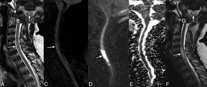
Evolution of ischemia. The first MR image shows the subtle signal anomaly on T2 and diffusion sequences (arrows, A–C). Follow-up 48 hours later shows an important tumefaction and high signal on T2WI associated with a restriction of diffusion of the cervical spinal cord at the C4–C7 levels (arrows, D–G).
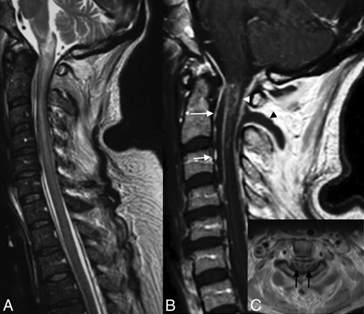
Venous infarction in a patient with epidural and paraspinal abscesses. Note large intramedullary high signal on T2 of the cervical spinal cord (A). T1WI with contrast medium demonstrates an intramedullary enhancement (B and C), the anterior (arrows, B) and posterior epidural (white arrowhead, B), and paraspinal abscesses (black arrowhead, B). Note enhancement on axial T1 of both sides of the median line, reflecting venous ischemia.
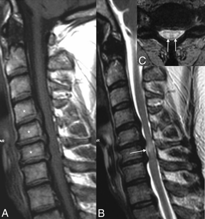
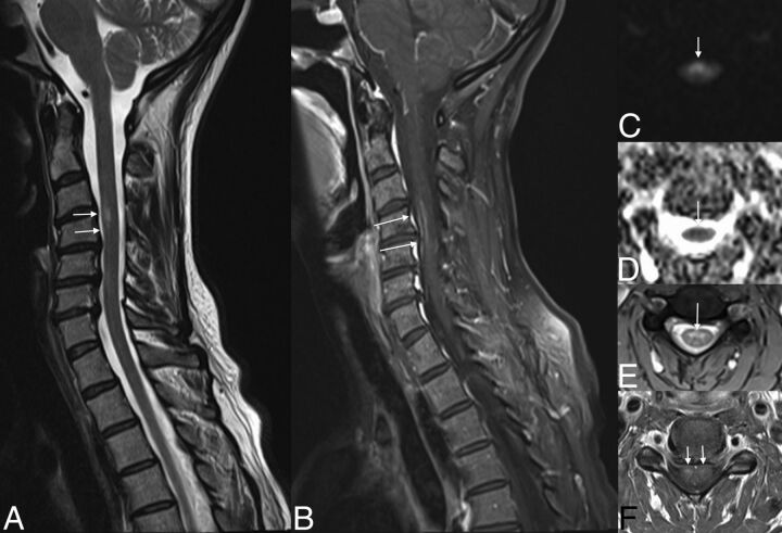
Cervical spinal canal stenosis and venous infarction. Note the cervical spinal canal stenosis from C4 to C6 due to cervical spondylosis (asterisks, A) and the intramedullary high signal on T2WI (arrow, B) at the same level with the “snake eye” appearance on axial T2WI (arrows, C).
Subacute ischemia. Note the slight hypersignal of the spinal anterior territory at the level of C4–C6 on T2WI (arrows, A and E), associated with a restriction of diffusion (arrows, C and D) and enhancement (arrows, D).
References
-
- Sandson TA, Friedman JH. Spinal cord infarction: report of 8 cases and review of the literature. Medicine 1989;68:282–92 - PubMed
-
- Piffaretti G, Bonardelli S, Bellosta R, et al. . Spinal cord ischemia after simultaneous and sequential treatment of multilevel aortic disease. J Thorac Cardiovasc Surg 2014;148:1435–42.e1 - PubMed
-
- Gorelik N, Tampieri D. Cocaine-induced vasospasm causing spinal cord transient ischemia. Neuroradiol J 2012;25:364–67 - PubMed
-
- Márquez JC, Granados AM, Castillo M. MRI of cervical spinal cord infarction in a patient with sickle cell disease. Clin Imaging 2012;36:595–98 - PubMed
Publication types
MeSH terms
LinkOut - more resources
Full Text Sources
Other Literature Sources