. 2015 Jan 15;118(2):212-23.
doi: 10.1152/japplphysiol.00463.2014. Epub 2014 Oct 16.

Cardiac troponin I Pro82Ser variant induces diastolic dysfunction, blunts β-adrenergic response, and impairs myofilament cooperativity

Affiliations

Cardiac troponin I Pro82Ser variant induces diastolic dysfunction, blunts β-adrenergic response, and impairs myofilament cooperativity

Genaro A Ramirez-Correa et al. J Appl Physiol (1985). .

Abstract

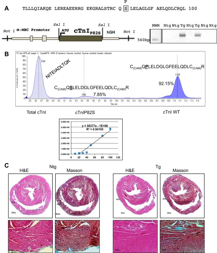
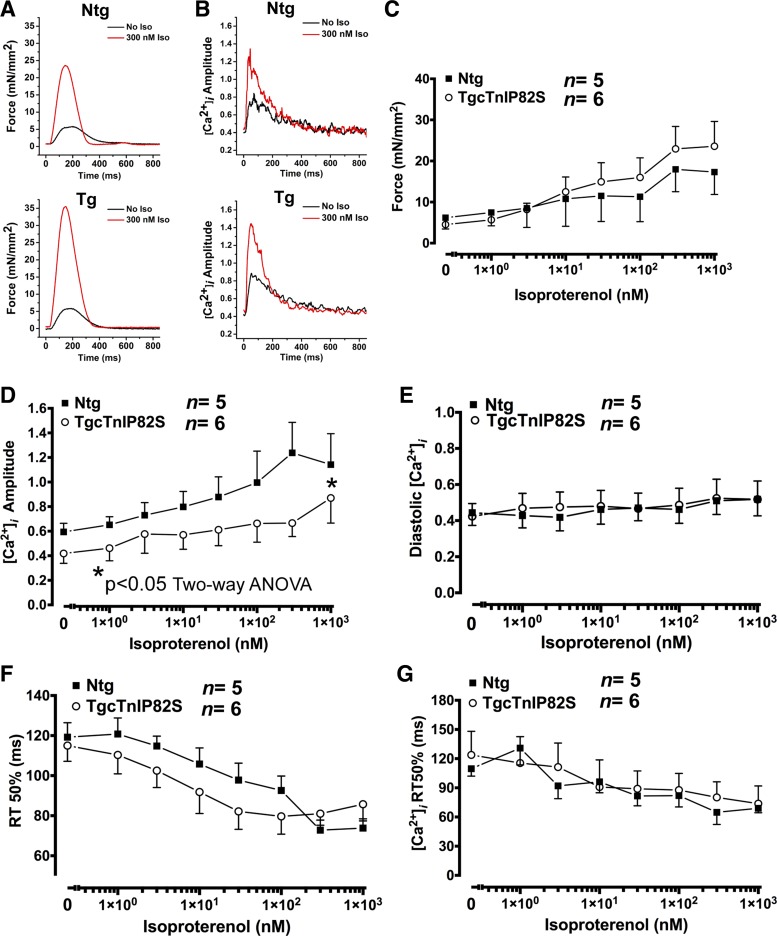
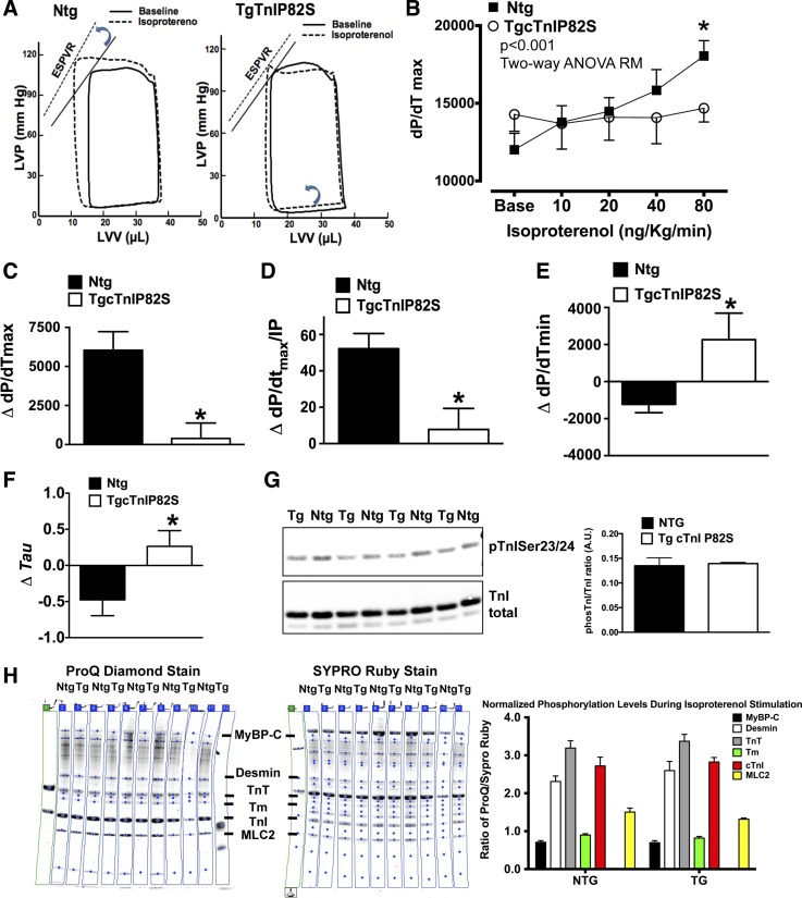
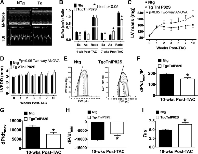
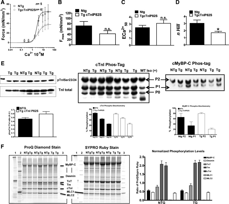
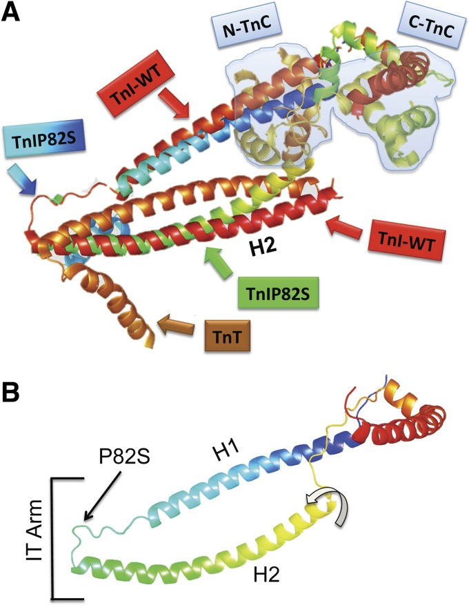
Troponin I (TnI) variant Pro82Ser (cTnIP82S) was initially considered a disease-causing mutation; however, later studies suggested the contrary. We tested the hypothesis of whether a causal link exists between cTnIP82S and cardiac structural and functional remodeling, such as during aging or chronic pressure overload. A cardiac-specific transgenic (Tg) mouse model of cTnIP82S was created to test this hypothesis. During aging, Tg cTnIP82S displayed diastolic dysfunction, characterized by longer isovolumetric relaxation time, and impaired ejection and relaxation time. In young, Tg mice in vivo pressure-volume loops and intact trabecular preparations revealed normal cardiac contractility at baseline. However, upon β-adrenergic stimulation, a blunted contractile reserve and no hastening in left ventricle relaxation were evident in vivo, whereas, in isolated muscles, Ca(2+) transient amplitude isoproterenol dose-response was blunted. In addition, when exposed to chronic pressure overload, Tg mice show exacerbated hypertrophy and decreased contractility compared with age-matched non-Tg littermates. At the molecular level, this mutation significantly impairs myofilament cooperative activation. Importantly, this occurs in the absence of alterations in TnI or myosin-binding protein C phosphorylation. The cTnIP82S variant occurs near a region of interactions with troponin T; therefore, structural changes in this region could explain its meaningful effects on myofilament cooperativity. Our data indicate that cTnIP82S mutation modifies age-dependent diastolic dysfunction and impairs overall contractility after β-adrenergic stimulation or chronic pressure overload. Thus cTnIP82S variant should be regarded as a disease-modifying factor for dysfunction and adverse remodeling with aging and chronic pressure overload.

Keywords: cardiac troponin I mutation; diastolic dysfunction; hypertrophy; transgenic mouse.


Figures

Fig. 1.
Fig. 1.
TgcTnIP82S mouse model displays normal histology. A: a fragment of cardiac troponin I (cTnI) cDNA where the mutation was introduced is shown in a gray shaded box. This mutant was cloned into a Sal I site downstream murine α-myosin heavy chain (MHC) promoter. Right: DNA 1% agarose gel showing two PCR products using primers specific for transgenic (Tg) mice illustrates two founders, from left to right, 270 and 273, positive genotype. B, top: a representative total ion chromatogram from a multiple reaction monitoring (MRM) assay quantifying the relative content of wild-type (WT) cTnI and cTnI P82S peptides with respect to total cTnI. Bottom: a representative calibration curve for mutant cTnIP82S peptide. A serial dilutions of mutant peptide were used to produce a 7-point calibration curve at 10, 20, 30, 40, 50, 80, and 100 pmol/μl for mutant P82S peptide [C(CAM)QSLELDGLGFEELQDLC(CAM)R̂]. C: TgcTnIP82S normal histology in representative hematoxylin and eosin (H&E), and lack of fibrosis in Masson's staining (×40 top row, and ×100 bottom row). Ntg, nontransgenic.
Fig. 2.
Fig. 2.
Intact twitching cardiac muscle shows blunted β-adrenergic dependent increase of Ca2+ transient amplitude. Representative twitch force (A) and calcium transients tracings (B) of Ntg and Tg, during isoproterenol (Iso) dose response at baseline (black) and 300 nM (red). C: developed force in response to increased dose of Iso (0 to 103 nM) for Ntg (solid squares) and Tg (open circles) isolated cardiac muscles. D: systolic Ca2+ transients amplitude (fura 2-AM, F1/F0) were significantly decreased in Tg, despite β-adrenergic stimulation. *P < 0.05, two-way ANOVA. E: diastolic Ca2+ transients in response to Iso. F: corresponding acceleration of force relaxation time (RT) to 50%. G: Ca2+ transient acceleration of decay to 50%. Values are means ± SE. [Ca2+]i, intracellular Ca2+ transients.
Fig. 3.
Fig. 3.
In vivo β-adrenergic dose-response is blunted in TgcTnIP82S. A: representative Ntg (n = 6) and Tg (n = 5) pressure-volume (PV) loops at baseline (solid line) and at highest point of Iso dose-response (10, 20, 40, and 80 ng·kg−1·min−1) (dashed line). Notice on left, a leftward and upward shift of Ntg PV loop, typical of adequate contractile adrenergic response, whereas the right shows the failure of TgcTnIP82S to respond to β-adrenergic stimulation. LVP, left ventricular pressure; LVV, left ventricular volume; ESPVR, end-systolic PV relationship. B: mean results for Iso dose-response of maximal rate constant of pressure rise (dP/dtmax). TgcTnIP82S mice show a blunted dose-dependent augmentation of systolic function vs. Ntg. *P < 0.001 is for group interaction terms analysis by two-way ANOVA repeated measures (RM). C: change (Δ) in dP/dtmax. D: Δmaximal rate of pressure rise normalized to instantaneous pressure (dP/dtmax/IP). E: Δpeak negative dP/dt (dP/dtmin). F: Δisovolumetric relaxation constant (τ), which was calculated by logistic regression. G: myofilament from Ntg and TgcTnIP82S that were subject to Iso dose-response showed no difference in TnI phosphorylation of Ser23/24. H: myofilament preparations from Ntg and TgcTnIP82S that were subject to Iso dose-response were determined by Pro-Q staining (left) and normalized to total protein content, determined by Sypro Ruby (middle). Comparison of normalized phosphorylation levels expressed as ratio of ProQ/Sypro Ruby signals. Phosphorylaton pattern is not significantly different (right). MyBP-C, myosin-binding protein C; MLC, myosin light chain. Values are means ± SE.
Fig. 4.
Fig. 4.
cTnIP82S exacerbates hypertrophic response and decreases contractility during chronic pressure-overload. A: representative M-mode and tissue Doppler imaging (TDI) echocardiograms of Ntg and Tg 1-wk post-transverse aortic constriction (TAC). B: reduced transmitral flow dynamics are evident in Tg mice (*t-test, P < 0.05). Ea, early diastolic myocardial velocity; Aa, late diastolic myocardial velocity. Left ventricular (LV) mass (C) and LV chamber dilatation (D) are significantly influenced by Tg genotype (*two-way ANOVA, P < 0.05). LVEDD, left ventricular end diastolic dimension. E: representative PV loops of Ntg and Tg 10 wk post-TAC at baseline (dashed line) and after imposing afterload (dashed line), respectively. Note that, although there is a right and upward shift of PV loop (contrary to a typical adequate contractility response), Ntg mice (n = 6) have overall better baseline and afterload contractility. This is clearly illustrated in FI, showing PV loop parameters that were significantly changed in Tg mice (n = 6). F: dP/dtmax/IP. G: dP/dtmax. H: dP/dtmin. I: τ. *P < 0.05. Values are means ± SE.
Fig. 5.
Fig. 5.
Steady-state force-Ca2+ relationships in skinned fibers. A: pool raw force data averaged and fitted to modified Hill equation: Ntg (n = 5), Tg (n = 6). B: averaged maximal Ca2+ activated force (Fmax). Note the trend of Tg to reduced Fmax. ns, Nonsignificant. C: Ca2+ sensitivity of Tg is modestly increased. ECa502+, extracellular Ca2+ concentration required for 50% of maximal activation D: myofilament cooperativity (n Hill, Hill coefficient) is markedly reduced in Tg muscles. *P < 0.05. E, left: TnI phosphorylation on Ser23/24 is not affected. PhosTag-SDS-PAGE show that phosphorylation stoichometry is unchanged for TnI (middle) and MyBP-C (right). F: phosphorylation of myofilament preparations from Ntg and TgcTnIP82S were determined by Pro-Q staining (left) and normalized to total protein content, determined by Sypro Ruby (middle). Molecular weight markers are labeled 1 (Precision-C, Bio-Rad), 2 (Peppermint Stick, Inv), and 3 (MagicMark XP, Inv) with myofilament protein migration highlighted by a line. Right: comparison of normalized phosphorylation levels expressed as ratio of ProQ/Sypro Ruby signals. Phosphorylation pattern is not significantly different. Values are means ± SE.
Fig. 6.
Fig. 6.
Molecular modeling of TnIP82S mutant. A: TnI WT and a structural model of TnIP82S are overimposed, maintaining the relation with TnT and TnC (light blue shaded). Cyan to blue gradient box and arrow show the proline → serine substitution. B: TnI P82S structural model and the functional domains. The hinge region of IT arm is where H1 and H2 join. Curved arrow shows the twist of H2 region would be further apart from TnT. To render the molecular models of TnI WT (PDB ID: 1J1D) and TnIP82S, we used the “backrub” method for flexible protein backbone modeling implemented in Rosetta.

References

    1. Biesiadecki BJ, Schneider KL, Yu ZB, Chong SM, Jin JP. An R111C polymorphism in wild turkey cardiac troponin I accompanying the dilated cardiomyopathy-related abnormal splicing variant of cardiac troponin T with potentially compensatory effects. J Biol Chem 279: 13825–13832, 2004. - PubMed
    1. Bilchick KC, Duncan JG, Ravi R, Takimoto E, Champion HC, Gao WD, Stull LB, Kass DA, Murphy AM. Heart failure-associated alterations in troponin I phosphorylation impair ventricular relaxation-afterload and force-frequency responses and systolic function. Am J Physiol Heart Circ Physiol 292: H318–H325, 2007. - PubMed
    1. Blanchard E, Seidman C, Seidman JG, LeWinter M, Maughan D. Altered crossbridge kinetics in the αMHC(403/+) mouse model of familial hypertrophic cardiomyopathy. Circ Res 84: 475–483, 1999. - PubMed
    1. Brown AT, Frazier A, Dennison CR, Hill MN, Post WS, Robinson JC, Murphy AM. A Non-synonymous variant of the cardiac troponin I gene is associated with enhanced cardiac hypertrophy response to hypertension in young black men (Abstract). In: AHA Scientific Basis of Heart Failure in Children. Dallas, TX: American Heart Association, 2008.
    1. Cardim N, Perrot A, Ferreira T, Pereira A, Osterziel KJ, Reis RP, Correia JF. Usefulness of Doppler myocardial imaging for identification of mutation carriers of familial hypertrophic cardiomyopathy. Am J Cardiol 90: 128–132, 2002. - PubMed

Publication types