. 2014 Nov 4;111(44):15768-73.
doi: 10.1073/pnas.1417518111. Epub 2014 Oct 16.

A nontranscriptional role for Oct4 in the regulation of mitotic entry

Affiliations

A nontranscriptional role for Oct4 in the regulation of mitotic entry

Rui Zhao et al. Proc Natl Acad Sci U S A. .

Abstract

Rapid progression through the cell cycle and a very short G1 phase are defining characteristics of embryonic stem cells. This distinct cell cycle is driven by a positive feedback loop involving Rb inactivation and reduced oscillations of cyclins and cyclin-dependent kinase (Cdk) activity. In this setting, we inquired how ES cells avoid the potentially deleterious consequences of premature mitotic entry. We found that the pluripotency transcription factor Oct4 (octamer-binding transcription factor 4) plays an unappreciated role in the ES cell cycle by forming a complex with cyclin-Cdk1 and inhibiting Cdk1 activation. Ectopic expression of Oct4 or a mutant lacking transcriptional activity recapitulated delayed mitotic entry in HeLa cells. Reduction of Oct4 levels in ES cells accelerated G2 progression, which led to increased chromosomal missegregation and apoptosis. Our data demonstrate an unexpected nontranscriptional function of Oct4 in the regulation of mitotic entry.

Keywords: CDC25; Cdk1; Oct4; mitotic entry; pluripotent stem cells.


Conflict of interest statement

Conflict of interest statement: G.Q.D. is a member of the scientific advisory boards of and holds stock in or receives consulting fees from the following companies: Johnson & Johnson, Verastem, Epizyme, iPierian, Solasia KK, and MPM Capital, LLP.

Figures

Fig. 1.
Fig. 1.
Oct4 forms a complex with Cdk1. (A) Coprecipitation of Cdk1 and cyclin B and A by streptavidin-agarose (SA) bead pulldown from the BirA-bioOct4 cell extracts. BirA cell extracts were used as controls. Arrowhead, phosphorylated Cdk1. (B) Coprecipitation of Oct4 by anti-Cdk1 antibody. Mock immunoprecipitation (IP) was performed using total IgG. (C) Coprecipitation of Oct4 and Cdk1 by Ni-NTA bead pulldown. Recombinant His6-cyclin A (Left) or His6-cyclin B (Right) was bound to Ni-NTA beads and incubated with ES-cell extracts. (D) Schematic of His6-tagged Oct4 mutants. N, N-terminal region; POUs, POU-specific domain; Homeo, Homeo domain; C, C-terminal region. (E) Domain mapping by His6-tagged Oct4 mutants. Recombinant His6-tagged full-length or truncated Oct4 were bound to Ni-NTA beads and then added to HeLa cell extracts. Shown are immunoblots of the indicated proteins after precipitation.
Fig. 2.
Fig. 2.
Oct4 inhibits Cdk1 activation in a cell extract-based kinase assay. (A) Representative immunoblots for Oct4 (Top) and Cdk1 (Bottom) after Oct4 immunodepletion and recombinant His6-Oct4 add-back. Arrow, His6-Oct4. (B) Representative autoradiogram of Cdk1 kinase assay in ES-cell extracts by adding increasing amounts of cyclin B after mock depletion, Oct4 depletion, or Oct4 depletion with His6-Oct4 add-back. (C) Quantification of three independent Cdk1 kinase assays in ES-cell extracts. Error bar, SEM. (D) Representative autoradiogram of Cdk1 kinase assay in HeLa-cell extracts with 2,000 nM cyclin B by adding increasing amounts of His6-Oct4. (E) Quantification of four independent Cdk1 kinase assays in HeLa-cell extracts. Error bar, SEM. (F) Representative autoradiogram of Cdk1 kinase assay in HeLa-cell extracts with 2,000 nM cyclin B and 2,000 nM Oct4. Oct4 was added with cyclin B or 20 min later. (G) Quantification of F (n = 3–6). Error bar, SEM. (H) Representative immunoblots for indicated proteins during the experiment shown in F. Arrowheads, phosphorylated proteins.
Fig. 3.
Fig. 3.
Oct4 inhibits Cdk1 activation in a recombinant protein-only Cdk1 kinase assay. (A) Coomassie-stained gel (Top) and autoradiogram (Bottom) of a representative cyclin B–Cdk1 kinase assay. The amounts of cyclin B–Cdk1 and CDC25C were too low for detection by Coomassie stain. Arrow represents position of mutant Oct4. (B) Quantification of nine independent kinase assays. Error bar, SEM; *P < 0.05; **P < 0.01.
Fig. 4.
Fig. 4.
Ectopic Oct4 expression in HeLa cells delays mitotic entry. (A) Schematic of the GFP-PCNA and H2B-RFP dual color system. (B) S-phase progression monitored by the GFP-PCNA reporter. (C) G2 phase is indirectly monitored by dispersal of the GFP-PCNA foci and condensation of H2B-RFP marked chromosomes. (D) M-phase progression monitored by the H2B-RFP reporter. (E) Immunoblots on HeLa control, HeLa cells expressing cyclin A (HeLa-cycA), Oct4 (HeLa-Oct4), and both (HeLa-Oct4 cycA) with indicated antibodies. (F) Timing of mitotic entry of HeLa control, HeLa-Oct4, HeLa-cycA, and HeLa-Oct4 cycA cells. These data represent three independent experiments (50 cells for each cell line counted per experiment). Error bar, SEM.
Fig. 5.
Fig. 5.
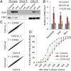
Oct4-mediated mitotic entry delay is transcription-independent. (A) Coprecipitation of Oct4 from HeLa cells expressing native Oct4 (wt) or a transcriptionally inactive mutant (mut) with Cdk1 antibody. Shown are immunoblots of indicated proteins. Mock IP was performed using total IgG. (B) Q-PCR analyses on Oct4 target genes in mouse embryonic fibroblasts (MEFs), MEFs expressing the native Oct4 (Oct4-wt) and the mutant Oct4 (Oct4-mut). Genes presented were induced reproducibly by Oct4-wt in three experiments. (C) Genome-wide expression profiling on HeLa control, HeLa Oct4-wt, and HeLa Oct4-mut cells. Presented are the Pearson’s correlation coefficient (r2) between two biological replicates of HeLa control cells (Top), HeLa control and HeLa Oct4-wt cells (Middle), and HeLa control and HeLa Oct4-mut cells (Bottom). (D) Timing of mitotic entry of HeLa control, HeLa Oct4-wt, and HeLa Oct4-mut cells. These data represent three independent experiments (50 cells for each cell line counted per experiment). Error bar, SEM.
Fig. 6.
Fig. 6.
Oct4 elongates G2 phase in ES cells. (A) Oct4 down-regulation in ZHBTc4 cells. Samples were collected at indicated time points with 10 μg/mL doxycycline treatment. Shown are immunoblots for indicated proteins. (B) FACS analysis of Oct4 expression level in ES cells. (C) FACS analysis of Oct4 down-regulated ES cells. ZHBTc4 cells treated with doxycycline for 12 h were stained for EdU incorporation and Oct4, Nanog, and DNA content. The cells were gated by Oct4 level into Oct4high, Oct4medium, and Oct4low populations (∼20% each of total Nanog-expressing cells). Cells negative for Nanog and expressing very low levels of Oct4 were excluded from the analysis. (D) Oct4 levels after release of ES cells from M-phase arrest. Shown are immunoblots for indicated proteins.
Fig. 7.
Fig. 7.
Oct4 helps maintain genomic integrity. (A) A representative image of micronuclei observed in the cytokinesis-block micronucleus assay in ZHBTc4 ES cells. Arrowheads, micronuclei enclosed in a telophase-arrested binuclei cell. (Left) DAPI staining; (Middle) bright field; (Right) merged. (Scale bar: 10 μm.) (B) Frequencies of micronuclei in control (dox−) and ZHBTc4 cells treated with doxycycline for 16 h (dox+) from four experiments. Error bar, SEM; two-tailed t test, **P < 0.01. (C) Percentage of Annexin-positive ZHBTc4 cells treated with doxycycline and DMSO or 9 μM RO-3306 for 16 h from five experiments. Error bar, SEM; *P < 0.05. (D) A model of Oct4 in the regulation of ES cell mitotic entry. (Top) In ES cells, a feedback loop due to inactivation of Rb and high levels of cyclins and Cdk activities creates the potential risk for premature mitotic entry. Oct4 serves as an additional, ES cell-specific mechanism to inhibit Cdk1 activation by counteracting CDC25 function and ensure timely mitotic entry. (Bottom) In somatic cells, expression of cyclins and activities of Cdk and Rb are regulated by the cell cycle.

References

    1. Becker KA, et al. Self-renewal of human embryonic stem cells is supported by a shortened G1 cell cycle phase. J Cell Physiol. 2006;209(3):883–893. - PubMed
    1. Fluckiger AC, et al. Cell cycle features of primate embryonic stem cells. Stem Cells. 2006;24(3):547–556. - PMC - PubMed
    1. Savatier P, Huang S, Szekely L, Wiman KG, Samarut J. Contrasting patterns of retinoblastoma protein expression in mouse embryonic stem cells and embryonic fibroblasts. Oncogene. 1994;9(3):809–818. - PubMed
    1. Stead E, et al. Pluripotent cell division cycles are driven by ectopic Cdk2, cyclin A/E and E2F activities. Oncogene. 2002;21(54):8320–8333. - PubMed
    1. Dulić V, Lees E, Reed SI. Association of human cyclin E with a periodic G1-S phase protein kinase. Science. 1992;257(5078):1958–1961. - PubMed

Publication types