. 2014 Dec 12;289(50):34900-10.
doi: 10.1074/jbc.M114.586362. Epub 2014 Oct 16.

Conformational response to ligand binding in phosphomannomutase2: insights into inborn glycosylation disorder

Affiliations

Conformational response to ligand binding in phosphomannomutase2: insights into inborn glycosylation disorder

Giuseppina Andreotti et al. J Biol Chem. .

Abstract

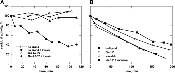
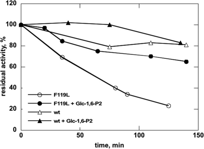
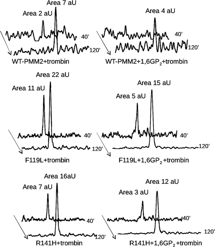
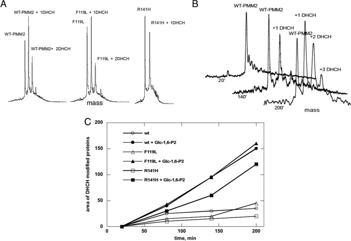
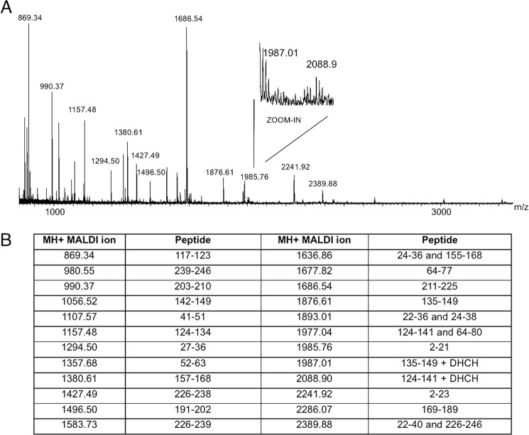
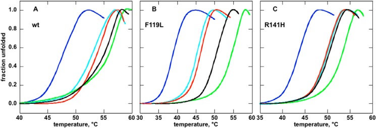
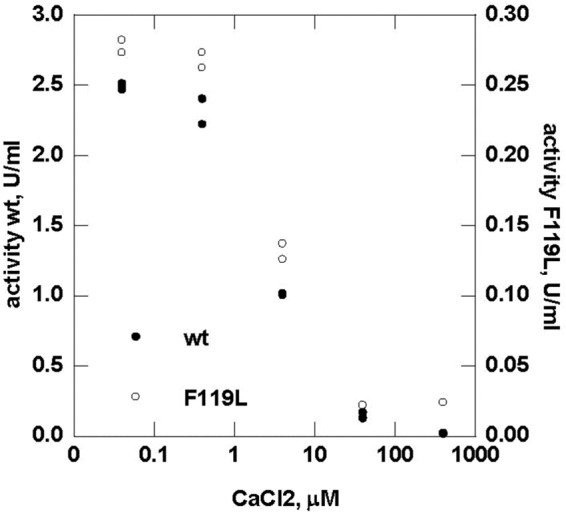
The most common glycosylation disorder is caused by mutations in the gene encoding phosphomannomutase2, producing a disease still without a cure. Phosphomannomutase2, a homodimer in which each chain is composed of two domains, requires a bisphosphate sugar (either mannose or glucose) as activator, opening a possible drug design path for therapeutic purposes. The crystal structure of human phosphomannomutase2, however, lacks bound substrate and a key active site loop. To speed up drug discovery, we present here the first structural model of a bisphosphate substrate bound to human phosphomannomutase2. Taking advantage of recent developments in all-atom simulation techniques in combination with limited and site-directed proteolysis, we demonstrated that α-glucose 1,6-bisphosphate can adopt two low energy orientations as required for catalysis. Upon ligand binding, the two domains come close, making the protein more compact, in analogy to the enzyme in the crystals from Leishmania mexicana. Moreover, proteolysis was also carried out on two common mutants, R141H and F119L. It was an unexpected finding that the mutant most frequently found in patients, R141H, although inactive, does bind α-glucose 1,6-bisphosphate and changes conformation.

Keywords: 1,6-Bisphosphate; Computer Modeling; Drug Discovery; Glycosylation; Glycosylation Inhibitor; Ligand-binding Protein; PELE; Phosphomannomutase.


Figures

FIGURE 1.
FIGURE 1.
Ligand binding energy profiles computed by PELE. A, binding energy profile in the full surface exploration against the ligand r.m.s.d. (heavy atom r.m.s.d.) to the bound reference structure. B, binding energy profile against the P-Mg distance along the ligand refinement process in the active site. In both panels, each dot corresponds to a binding energy (see “Experimental Procedures”) obtained from a protein-ligand complex obtained in PELE sampling. Different colors correspond to processors.
FIGURE 2.
FIGURE 2.
Protein closure along ligand binding. The initial model in open conformation is shown in pale blue, and the model in closed conformation is shown in dark blue. Side chains of two residues that come close upon domain closure, Arg21 and Gln138, are shown as sticks. The initial and final positions of the ligand are shown in pink and red, respectively, with a ball and stick representation. The images were drawn with Chimera (27).
FIGURE 3.
FIGURE 3.
Ligand binding interactions. Shown is the active site protein-ligand interaction scheme for the P-Mg and P′-Mg binding modes obtained along the refinement sampling. The structures can be downloaded from the supplemental information.
FIGURE 4.
FIGURE 4.
PMM2, PMM1, and PMM_LEIME sequence alignment. Sequence alignment of PMM2, PMM1, and PMM_LEIME is shown. Active site residues are highlighted. The alignment shows that active site residues are less conserved between the two paralogous human enzymes than between PMM2 and the protozoan enzyme ortholog.
FIGURE 5.
FIGURE 5.
Limited proteolysis of wild type phosphomannomutase2 with trypsin monitored by assaying residual mutase activity. Purified wild type phosphomannomutase2 (1.4 mg/ml in 20 mm Hepes, pH 7.5 containing 150 mm NaCl and 1 mm MgCl2) was incubated at 37 °C with (or without) trypsin (protease:enzyme ratio, 1:20) in the presence of 0.5 mm Glc-1,6-P2 (A) or 0.5 mm Glc-1-P, 0.5 mm vanadate, or 0.5 mm Glc-1-P plus 0.5 mm vanadate (B). Residual phosphomannomutase activity is reported as a percentage with activity at time 0 defined as 100%. The protective effect of ligands increases as negative charges are added.
FIGURE 6.
FIGURE 6.
Wild type and F119L proteolysis with thrombin monitored by assaying residual mutase activity. Purified wild type or F119L phosphomannomutase2 (0.4 mg/ml in 20 mm Hepes, pH 7.5 containing 150 mm NaCl and 1 mm MgCl2) was incubated at 37 °C with thrombin (500 milliunits/μg of protein) with or without 0.5 mm Glc-1,6-P2. Residual phosphoglucomutase is reported as a percentage with activity at time 0 defined as 100%. Aliquots were also analyzed by mass spectrometry.
FIGURE 7.
FIGURE 7.
Limited proteolysis of wild type, F119L, or R141H phosphomannomutase2 with thrombin monitored by mass spectrometry. The extracted ion chromatograms relative to the formation of peptide 2–21 are reported for wild type (A and B), F119L (C and D), and R141H (E and F) phosphomannomutase2. Samples were incubated in the absence (A, C, and E) or presence (B, D, and F) of α-Glc-1,6-P2 (1,6GP2) with thrombin for 40 or 120 min. Peak areas were calculated by MassLynx 4.0 software and are reported as arbitrary units (aU).
FIGURE 8.
FIGURE 8.
Identification of the peptide generated by thrombin hydrolysis of phosphomanomutase2. The electrospray ionization-MS-MS spectrum of the peak at m/z 993.0, corresponding to the doubly charged ion of 1984.0 Da attributed to peptide 2–21, shows two major fragments that are compatible with the loss of Ala-Ala from the N terminus (doubly charged y-series fragment at 922.53) and with the loss of Leu-Thr-Ala-Pro-Arg from the C terminus (doubly charged b-series fragment at 715.47).
FIGURE 9.
FIGURE 9.
Chemical modification of reactive arginines in phosphomannomutase2 monitored by mass spectrometry. A shows deconvoluted RP-LC-MS mass spectra of WT-PMM2, F119L, and R141H incubated with 1,2-cyclohexanedione (molar excess of 100) for 140 min, revealing the different modification levels of the three proteins. B shows the time course modification of WT-PMM2, revealing unmodified protein (molecular mass of 27,950.25 Da) and mono- and di-modified species (molecular masses of 28,061.50 and 28,173.10 Da). C shows the graph of the area of the peaks of deconvoluted spectra relative to 1,2-cyclohexanedione-modified WT-PMM2, F119L, and R141H with or without Glc-1,6-P2 measured at different incubation times. ′, minutes.
FIGURE 10.
FIGURE 10.
Identification of reactive arginines in phosphomannomutase2 monitored by MALDI mass spectrometry. A shows the MALDI mass spectrum of WT-PMM2 incubated with 1,2-cyclohexanedione (molar excess of 100) for 140 min and digested by trypsin for 90 min. B shows the list of MH+ values measured by MALDI MS and attributed to tryptic peptides. Values at 1987.01 and 2088.90 atomic mass units were compatible with peptides 135–149 and 124–141 alternatively modified by 1,2-cyclohexanedione at Arg141 and Arg134, respectively.
FIGURE 11.
FIGURE 11.
Thermal stability of wild type, F119L, or R141H phosphomannomutase2 monitored by thermal shift assay. The profiles (A, WT; B, F119L; C, R141H) were assessed by thermal shift assays. Proteins (0.275 mg/ml in 20 mm Hepes, pH 7.5, 2 mm MgCl2, 150 mm NaCl, 1 mm dithiothreitol, 2.4× SYPRO Orange) were heated from 25 to 80 °C at 0.5 °C/min, and fluorescence was recorded (excitation, 490 nm; emission, 575 nm). The experiment was performed in the presence of no ligand (light blue), 5 mm EDTA (blue), 0.5 mm Glc-1-P (red), 0.5 mm Glc-1-P plus 0.5 mm vanadate (black), and 0.5 mm Glc-1,6-P2 (green).
FIGURE 12.
FIGURE 12.
Effect of calcium ions on the enzymatic activity of wild type and F119L phosphomannomutase2. Phosphomannomutase activity of wild type and F119L was measured in the presence of calcium chloride ranging from 0 to 400 μm.

References

    1. Matthijs G., Schollen E., Heykants L., Grünewald S. (1999) Phosphomannomutase deficiency: the molecular basis of the classical Jaeken syndrome (CDGS type Ia). Mol. Genet. Metab. 68, 220–226 - PubMed
    1. Stenson P. D., Mort M., Ball E. V., Shaw K., Phillips A., Cooper D. N. (2014) The Human Gene Mutation Database: building a comprehensive mutation repository for clinical and molecular genetics, diagnostic testing and personalized genomic medicine. Hum. Genet. 133, 1–9 - PMC - PubMed
    1. Schollen E., Kjaergaard S., Legius E., Schwartz M., Matthijs G. (2000) Lack of Hardy-Weinberg equilibrium for the most prevalent PMM2 mutation in CDG-Ia (congenital disorders of glycosylation type Ia). Eur. J. Hum. Genet. 8, 367–371 - PubMed
    1. Kjaergaard S., Skovby F., Schwartz M. (1998) Absence of homozygosity for predominant mutations in PMM2 in Danish patients with carbohydrate-deficient glycoprotein syndrome type 1. Eur. J. Hum. Genet. 6, 331–336 - PubMed
    1. Bjursell C., Wahlström J., Berg K., Stibler H., Kristiansson B., Matthijs G., Martinsson T. (1998) Detailed mapping of the phosphomannomutase 2 (PMM2) gene and mutation detection enable improved analysis for Scandinavian CDG type I families. Eur. J. Hum. Genet. 6, 603–611 - PubMed

Publication types

MeSH terms

LinkOut - more resources