Clinical Trial
. 2014 Nov 28;289(48):33391-403.
doi: 10.1074/jbc.M114.579870. Epub 2014 Oct 16.

Asbestos-induced disruption of calcium homeostasis induces endoplasmic reticulum stress in macrophages

Affiliations
Clinical Trial

Asbestos-induced disruption of calcium homeostasis induces endoplasmic reticulum stress in macrophages

Alan J Ryan et al. J Biol Chem. .

Abstract

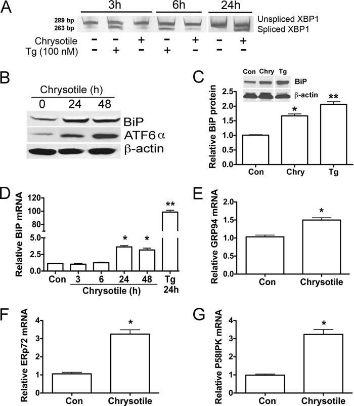
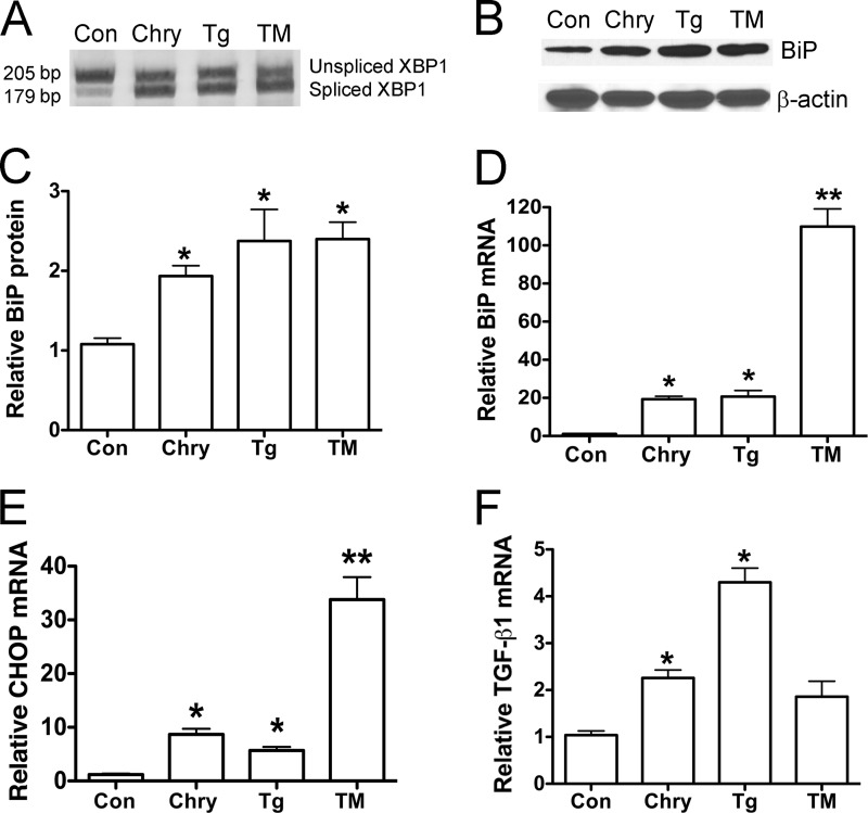
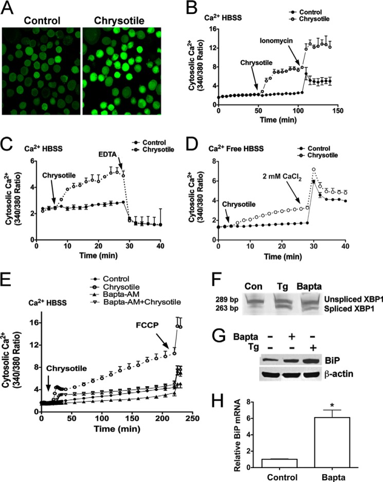
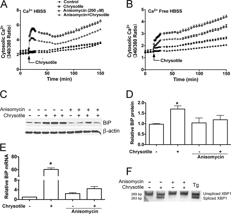
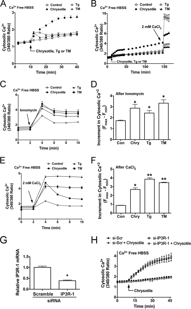
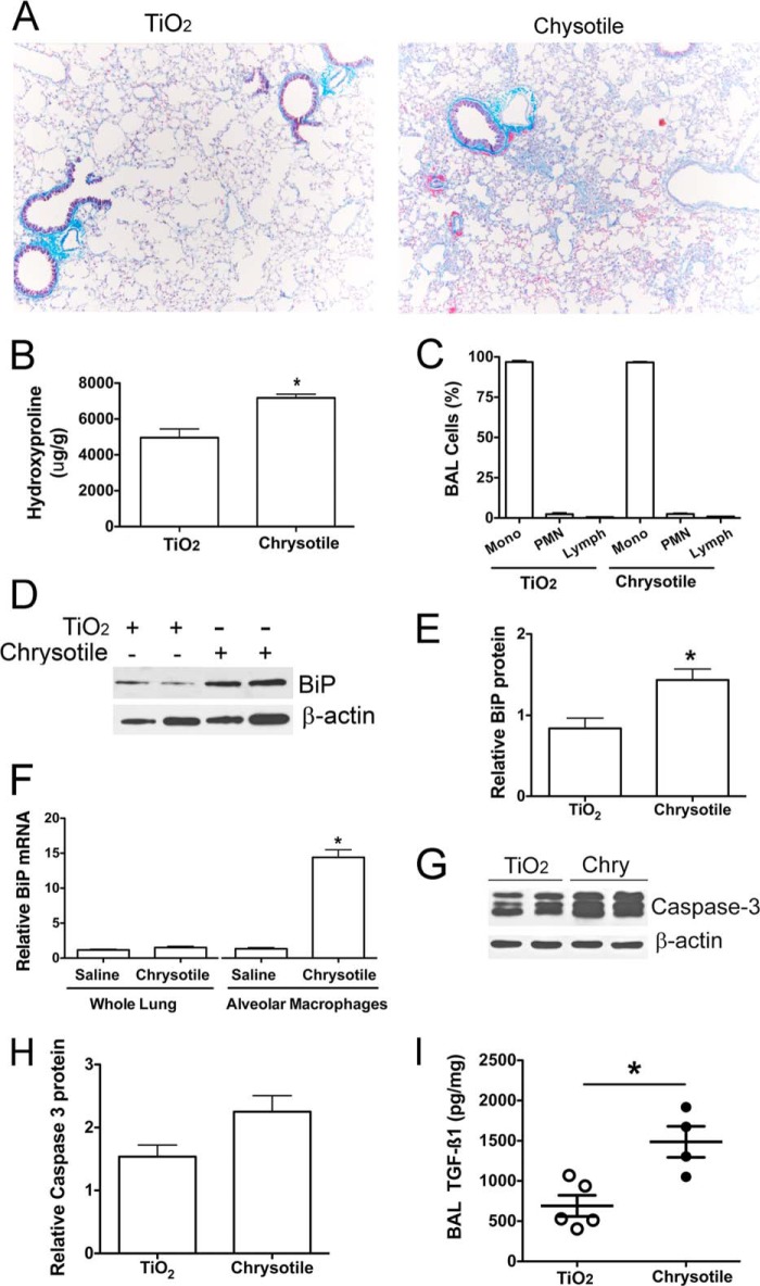
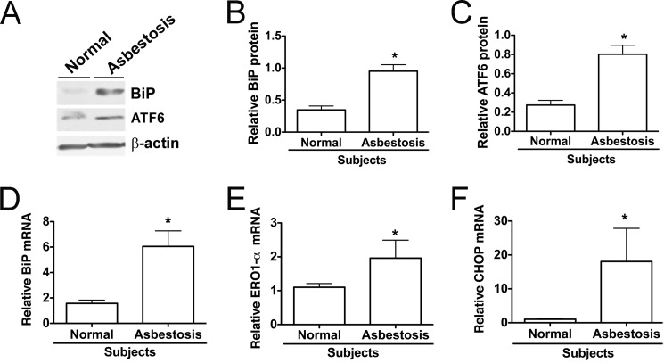
Although the mechanisms for fibrosis development remain largely unknown, recent evidence indicates that endoplasmic reticulum (ER) stress and activation of the unfolded protein response (UPR) may act as an important fibrotic stimulus in diseased lungs. ER stress is observed in lungs of patients with idiopathic pulmonary fibrosis. In this study we evaluated if ER stress and the UPR was present in macrophages exposed to chrysotile asbestos and if ER stress in macrophages was associated with asbestos-induced pulmonary fibrosis. Macrophages exposed to chrysotile had elevated transcript levels of several ER stress genes. Macrophages loaded with the Ca(2+)-sensitive dye Fura2-AM showed that cytosolic Ca(2+) increased significantly within minutes after chrysotile exposure and remained elevated for a prolonged time. Chrysotile-induced increases in cytosolic Ca(2+) were partially inhibited by either anisomycin, an inhibitor of passive Ca(2+) leak from the ER, or 1,2-bis(2-aminophenoxyl)ethane-N,N,N',N'-tetraacetic acid (BAPTA-AM), an intracellular Ca(2+) chelator known to deplete ER Ca(2+) stores. Anisomycin inhibited X-box-binding protein 1 (XBP1) mRNA splicing and reduced immunoglobulin-binding protein (BiP) levels, whereas BAPTA-AM increased XBP1 splicing and BiP expression, suggesting that ER calcium depletion may be one factor contributing to ER stress in cells exposed to chrysotile. To evaluate ER stress in vivo, asbestos-exposed mice showed fibrosis development, and alveolar macrophages from fibrotic mice showed increased expression of BiP. Bronchoalveolar macrophages from asbestosis patients showed increased expression of several ER stress genes compared with normal subjects. These findings suggest that alveolar macrophages undergo ER stress, which is associated with fibrosis development.

Keywords: Asbestos; Calcium; Endoplasmic Reticulum Stress (ER Stress); Fibrosis; Lung Injury; Macrophage; Pulmonary Fibrosis.


Figures

FIGURE 1.
FIGURE 1.
Chrysotile induces ER stress in macrophages. A, macrophages were exposed to thapsigargin (Tg, 100 nm) or chrysotile (10 μg/cm2) for the indicated times. Spliced XBP1 was evaluated by conventional RT-PCR followed by acrylamide gel electrophoresis. B, macrophages exposed to chrysotile (10 μg/cm2) for 24 or 48 h and cell lysates were used for GRP 78/BiP and ATF6 expression by protein immunoblot analysis. C, densitometric analysis of BiP protein expression corrected for β-actin in cells exposed to chrysotile (Chry) or thapsigargin for 24 h. * or **, p < 0.05 versus control or thapsigargin or Chry. n = 8/group. Inset shows an BiP immunoblot for a 24-h exposure. D, quantitative RT-PCR for BiP mRNA corrected for hypoxanthine-guanine phosphoribosyltransferase mRNA. Macrophages were treated with chrysotile for the indicated times or thapsigargin for 24 h. * or **, p < 0.05 versus other groups. n = 8/group. E–G, quantitative RT-PCR data showing mRNA of different ER stress genes corrected for hypoxanthine-guanine phosphoribosyltransferase mRNA. n = 3/group. RNA was isolated from macrophages 24 h after exposure to chrysotile (10 μg/cm2). *, p < 0.05 versus control. n = 3/group.
FIGURE 2.
FIGURE 2.
Chrysotile induces ER stress in bone marrow-derived macrophages. A, macrophages were exposed to chrysotile (10 μg/cm2), thapsigargin (Tg, 100 nm), or tunicamycin (TM, 5 μg/ml) for 24 h, and then assayed for XBP1 splicing using conventional RT-PCR plus acrylamide gel electrophoresis. Data from experiments conducted as described in A are shown: B, immunoblot analysis for BiP protein and β-actin; C, densitometric analysis of BiP protein expression was corrected for β-actin (*, p < 0.05 versus control). D–F, quantitative RT-PCR data showing relative BiP mRNA, relative CHOP mRNA, and relative TGFβ1 mRNA corrected β-actin. *, p < 0.05 versus control; **, p < 0.05 versus other groups. n = 6–10/group.
FIGURE 3.
FIGURE 3.
Chrysotile increases cytosolic Ca2+ levels in macrophages. A, cells were loaded with Fluo3-AM (2.5 μm, 1 h, 37 °C) and then exposed to chrysotile (10 μg/cm2) for evaluation by confocal microscopy. Cells were loaded with Fura2-AM (1 μm, 30 min, 37 °C), suspended in HBSS containing 1.3 mm Ca2+, and exposed to chrysotile (10 μg/well) followed by (B) 5 μm ionomycin or (C) 2 mm EDTA. Values are mean ± S.E. (n = 3/group). D, cells were loaded with Fura2-AM as above stated, suspended in Ca2+-free HBSS, exposed to chrysotile (10 μg/cm2) followed by 2 mm CaCl2. E, cells were loaded with Fura2-AM (1 μm, 30 min, 37 °C) and Ca2+ chelator BAPTA-AM (1 μm, 30 min, 37 °C) 30 min before chrysotile exposure. The proton ionophore FCCP (5 μm) was used as a positive control. n = 6/group. Cells were exposed to BAPTA-AM (5 μm, 24 h) and assayed for (F) spliced XBP1, by conventional RT-PCR plus gel electrophoresis as well as (G) BiP protein expression by immunoblot analysis, and (H) BiP mRNA expression by quantitative RT-PCR. Thapsigargin (Tg) (100 nm) for 24 h was used as a positive control. *, p < 0.05 versus control (n = 6/group).
FIGURE 4.
FIGURE 4.
Anisomycin treatment reduces cytosolic Ca2+ levels and modulates chrysotile-induced ER stress. Cells were loaded with Fura2-AM (1 μm, 30 min, 37 °C) and suspended in either HBSS containing 1.3 mm Ca2+ HBSS (A) or Ca2+ free HBSS (B). Cells were incubated with 200 μm anisomycin for 60 min before exposure to chrysotile (10 μg/cm2). Values are mean ± S.E. (n = 6/group). C, macrophages were pretreated with 0.2 μm anisomycin for 60 min before exposure to chrysotile (10 μg/cm2) for 24 h. BiP protein in lysates was determined by immunoblot analysis. Data from experiments conducted as described in C are shown: D, densitometric analysis of BiP protein corrected for β-actin (mean ± S.E., 3 experiments); E, BiP mRNA corrected for hypoxanthine-guanine phosphoribosyltransferase mRNA assayed by quantitative RT-PCR (*, p < 0.05 versus control, mean ± S.E., 3 experiments); and F, XBP1 splicing was evaluated by conventional RT-PCR followed by acrylamide gel electrophoresis.
FIGURE 5.
FIGURE 5.
Chrysotile increases ionomycin-releasable calcium stores and alters activation of store-operated Ca2+ channels in macrophages. A, cells were loaded with Fura2-AM (1 μm, 30 min, 37 °C), suspended in Ca2+-free HBSS, and exposed to chrysotile (10 μg/well), thapsigargin (Tg) (100 nm), or tunicamycin (TM) (5 μg/ml) followed by (B) 2 mm CaCl2. Values are mean ± S.E. (n = 6/group). C, cells were exposed to chrysotile (10 μg/well), thapsigargin (100 nm), or tunicamycin (5 μg/ml) for 24 h and then assayed for Ca2+ release by ionomycin (5 μm). D, increments in cytosolic Ca2+ were determined by the maximum change in F340/380 observed in the first 2 min after ionomycin. *, p < 0.05 versus control (n = 6/group). E, cells were exposed to chrysotile, thapsigargin, or tunicamycin for 24 h as described above, and then assayed for changes in cytosolic Ca2+ after addition of 2 mm CaCl2. F, increments in cytosolic Ca2+ after CaCl2 addition were determined as described in D. * or **, p < 0.05 versus control (n = 6/group). E, macrophages were transfected with either scrambled or IP3R-1 siRNA for 48 h and assayed for IP3R-1 mRNA by quantitative RT-PCR (*, p < 0.05 versus scramble) (G) and cytosolic Ca2+ levels after chrysotile exposure (H) (n = 6/group).
FIGURE 6.
FIGURE 6.
ER stress is present in macrophages obtained from fibrotic lungs. C57BL/6J mice were given either TiO2 (125 μg) or chrysotile (125 μg) intratracheally. BAL and lung tissues were obtained 21 days after exposure. A, lung sections from mice exposed to TiO2 or chrysotile were assayed by Masson's trichrome staining for detection of collagen deposition. B, hydroxyproline content of mouse lung exposed to TiO2 or chrysotile for 21 days. *, p < 0.05 versus TiO2 (n = 6/group). C, differential counts for BAL cells after Giemsa-Wright staining (n = 6/group). D, BAL cell lysates assayed by BiP protein immunoblotting. E, densitometric analysis of BiP protein expression corrected for β-actin; *, p < 0.05 versus TiO2 (n = 4/group). F, total RNA from whole lung or from BAL cells was evaluated by quantitative RT-PCR for BiP mRNA normalized to β-actin mRNA. *, p < 0.05 versus other groups (n = 3/group). G, BAL cell lysates were assayed by protein immunoblotting for caspase-3 and β-actin. H, densitometric analysis of caspase-3 protein corrected for β-actin (n = 4/group, p = 0.06). I, BAL fluid assayed for TGF-β1 by ELISA. Values normalized to BAL protein, n = 4–5/group.
FIGURE 7.
FIGURE 7.
Alveolar macrophages obtained from asbestosis patients show indicators of ER stress. A, alveolar macrophage lysates used for assay of BiP and ATF6 protein expression by immunoblotting. B and C, densitometric analysis of BiP and ATF6 protein expression normalized to β-actin in normal subjects and asbestosis patients. *, p < 0.05 versus normal subjects (n = 3–4/group). D–F, quantitative RT-PCR data showing mRNA of different ER stress genes corrected for hypoxanthine-guanine phosphoribosyltransferase mRNA. *, p < 0.05 versus normal subjects (n = 3/group).

References

    1. Guidotti T. L., Miller A., Christiani D., Wagner G., Balmes J., Harber P., Brodkin C. A., Rom W., Hillerdal G., Harbut M., Green F. H. Y. (2004) Diagnosis and initial management of nonmalignant diseases related to asbestos. Am. J. Respir. Crit. Care Med. 170, 691–715 - PubMed
    1. Lenna S., Trojanowska M. (2012) The role of endoplasmic reticulum stress and the unfolded protein response in fibrosis. Curr. Opin. Rheumatol. 24, 663–668 - PMC - PubMed
    1. Tanjore H., Blackwell T. S., Lawson W. E. (2012) Emerging evidence for endoplasmic reticulum stress in the pathogenesis of idiopathic pulmonary fibrosis. Am. J. Physiol. Lung Cell. Mol. Physiol. 302, L721–729 - PMC - PubMed
    1. Hetz C. (2012) The unfolded protein response: controlling cell fate decisions under ER stress and beyond. Nat. Rev. Mol. Cell Biol. 13, 89–102 - PubMed
    1. Kim I., Xu W., Reed J. C. (2008) Cell death and endoplasmic reticulum stress: disease relevance and therapeutic opportunities. Nat. Rev. Drug Discov. 7, 1013–1030 - PubMed

Publication types

MeSH terms