. 2014 Oct 2:8:303.
doi: 10.3389/fncel.2014.00303. eCollection 2014.

Maximum likelihood estimation of biophysical parameters of synaptic receptors from macroscopic currents

Affiliations

Maximum likelihood estimation of biophysical parameters of synaptic receptors from macroscopic currents

Andrey Stepanyuk et al. Front Cell Neurosci. .

Abstract

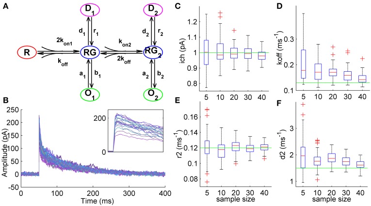
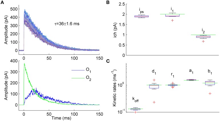
Dendritic integration and neuronal firing patterns strongly depend on biophysical properties of synaptic ligand-gated channels. However, precise estimation of biophysical parameters of these channels in their intrinsic environment is complicated and still unresolved problem. Here we describe a novel method based on a maximum likelihood approach that allows to estimate not only the unitary current of synaptic receptor channels but also their multiple conductance levels, kinetic constants, the number of receptors bound with a neurotransmitter, and the peak open probability from experimentally feasible number of postsynaptic currents. The new method also improves the accuracy of evaluation of unitary current as compared to the peak-scaled non-stationary fluctuation analysis, leading to a possibility to precisely estimate this important parameter from a few postsynaptic currents recorded in steady-state conditions. Estimation of unitary current with this method is robust even if postsynaptic currents are generated by receptors having different kinetic parameters, the case when peak-scaled non-stationary fluctuation analysis is not applicable. Thus, with the new method, routinely recorded postsynaptic currents could be used to study the properties of synaptic receptors in their native biochemical environment.

Keywords: Markov chain Monte Carlo; kinetic model; maximum likelihood; peak-scaled non-stationary fluctuation analysis; semiseparable matrix; synaptic currents; unitary current.


Figures

Figure 1
Figure 1
Estimation of unitary current and kinetic constants from simulated GABAergic synaptic currents. (A) 7-state kinetic scheme of GABAA receptor that was used to simulate macroscopic synaptic currents (Mozrzymas et al., , see Section Simulation of Macroscopic Synaptic Currents in Methods). The scheme has one unbound state, R, two liganded states (single-liganded, RG, and double-liganded, RG2,) and related open (O1 and O2) and desensitized (D1 and D2) states. Rate constants were adapted from Mozrzymas et al. (2003) and were as follows: koff = 0.13 ms−1, d1 = 0.14 ms−1, d2 = 1.5 ms−1, r1 = 0.02 ms−1, r2 = 0.12 ms−1, a1 = 1.5 ms−1, a2 = 1 ms−1, b1 = 0.15 ms−1, b2 = 8 ms−1; kon1 = 4 mM−1 ms−1, kon2 = 8 mM−1 ms−1. Unitary currents for the states O1 and O2 were equal and were set to i1 = i2 = 1 pA. The number of channels exposed to GABA varied from trial to trail (Nch = 250, SD = 50; Gaussian variation). Colored noise (SD = 3 pA) was added to the simulated currents (see Section Simulation of Macroscopic Synaptic Currents in Methods). (B) Synaptic currents simulated using the kinetic scheme shown in (A). The currents demostrate high trial-to-trial variability resembling one observed in experimental electrophysiological recordings (inset). (C) Statistical plots demonstrating accuracy of unitary current estimates obtained by ML NSFA. On each plot, the central mark (red) is the median, the edges of the box are the 25th and 75th percentiles, the whiskers extend to the most extreme data points not considered outliers, and outliers are plotted individually by red crosses. Green line indicates true value of unitary current (1 pA). Note high accuracy of unitary current estimates obtained by ML NSFA even if a few (5–20) currents were used. (D–F) Statistical plots of estimates of some kinetic constants obtained by ML NSFA. Colors are the same as in (C).
Figure 2
Figure 2
ML NSFA is more accurate than PS NSFA in estimating of unitary current. Estimation of the number of receptors bound with a neurotransmitter and peak open probability with ML NSFA. (A) Statistical plots demonstrating accuracy of unitary current estimates obtained with ML NSFA (blue boxes) and PS NSFA (black boxes) from simulated macroscopic synaptic currents with trial-to-trial Gaussian variation in the number of receptors (Nch = 250, SD = 50; see kinetic scheme in Figure 1A). On each plot, the central mark (red) is the median, the edges of the box are the 25th and 75th percentiles, the whiskers extend to the most extreme data points not considered outliers, and outliers are plotted individually by red crosses. Green line indicates a true value of unitary current. ML NSFA and PS NSFA were performed using n = 60 and n = 1000 samples consisting of N = 5, 10, 20, 30, 40 or 100 simulated currents, respectively. Note that the accuracy of estimates obtained with ML NSFA using a few (5–20) currents was 2-times better than one obtained with PS NSFA. (B) An example of variance vs. mean plot (gray dots) obtained with PS NSFA for N = 30 simulated macroscopic currents having trial-to-trial Gaussian variation in the number of receptors (Nch = 250, SD = 50) and a parabolic fit of its rising phase (red). Note that variance-mean relationship (gray dots) is skewed rather than parabolic and therefore the number of receptors could not be estimated with PS NSFA. (C,D) Statistical plots for the estimates of the number of channels bound with a neurotransmitter right after the concentration transient, Nch, and peak open probability, P(o, peak) obtained with ML NSFA. Green line in C indicated the true value of the number of channels estimated as mean peak current amplitude (averaged over N = 1000 currents) divided by the true value of P(o, Peak) and by the true value of unitary current (1 pA) and in D green line indicates the true value of P(o, Peak) estimated as P(o) = eQtp0. Other colors and notations are the same as in Figure 1C.
Figure 3
Figure 3
Estimation of unitary currents and kinetic constants of receptors having two open states with different conductance levels. (A) Upper panel. Example of 50 synaptic currents simulated with a 7-state kinetic scheme of GABAA receptor having two open states (Figure 1A, some rate constants were modified: b2 = 4, b1 = 1.2, d1 = 1, r1 = 1, d2 = 0.15, r2 = 1 ms−1). Unitary currents were set to i1 = 2 pA and i2 = 1 pA for open states O1 and O2, respectively. The number of channels varied from trial to trail (Nch = 500 ± 50; Gaussian variation). Lower panel. Representative example of single simulated macroscopic current components mediated by single-liganded open state O1 (blue trace) and double-liganded open state O2 (green trace) demonstrating comparable contribution of O1 and O2 to the total macroscopic current. (B) Statistical plots for the estimates of unitary currents obtained with PS NSFA (leftmost bar, i = 1.86 ± 0.03 pA) and ML NSFA (two bars on the right, i1 = 2.0 ± 0.11 pA and i2 = 0.89 ± 0.08 pA, i1 and i2 are unitary currents associated with open states O1 and O2, respectively. Both PS NSFA and ML NSFA were applied to samples of 50 macroscopic currents (n = 15 and n = 250 bootstrap samples for MS NSFA and PS NSFA, respectively) simulated as described in (A) and having true values of i1(0) = 2 pA and i2(0) = 1 pA, respectively (indicated by green lines). On each plot, the central mark (red) is the median, the edges of the box are the 25th and 75th percentiles, the whiskers extend to the most extreme data points not considered outliers, and outliers are plotted individually by red crosses. Note that ML NSFA accurately distinguishes both unitary current levels, whereas PS NSFA gave some value of the unitary current that was close to i1(0). (C) Statistical plot for the estimates of kinetic rates of transitions from and to a single-liganded state obtained by ML NSFA (in ms−1: unbinding rate, koff = 0.13 ± 0.01, desensitization rate, d1 = 0.89 ± 0.34, resensitization rate r1 = 1.02 ± 0.08, closing rate, a1 = 1.55 ± 0.05, opening rate, b1 = 1.17 ± 0.24; N = 50 currents simulated as described in (A). The estimates were in good agreement with their true values (green lines). See a legend to panel (B) for further description.
Figure 4
Figure 4
ML NSFA distinguishes between changes in the receptor gating and the number of receptors in case when both unitary current and macroscopic current waveform are not changed. (A) Simple 3-state kinetic scheme of synaptic receptor channel. The scheme consists of one unbound state, R, one single-liganded state, RL, and one open state, O. Rate constants are shown below the respective transitions and were as follows: kon = 6 mM−1 ms−1, koff = 0.025 ms−1, b = 0.25 ms−1. Three different models were constructed based on this scheme and were used to simulate 3 sets of macroscopic currents. A closing rate constant, a, was set to 2.5 ms−1 for Model R (red) and Model N (black) and 1.25 ms−1 for Model A (blue). (B) An example of 3 macroscopic currents simulated using Model R (red), Model A (blue), and Model N (black) shown in (A). The number of channels used for simulations is indicated in a respective color in the top-right corner (400 ± 50 for Models R and A and 800 ± 71 for Model N). (C) Mean simulated currents for each Model (N = 1000). Note that amplitudes of mean currents obtained with Model A and Model N (blue and black) are almost equal and almost twice larger than the mean current amplitude obtained with a reference Model R (red). (D) The same mean currents as in (C) but normalized. Note that all 3 waveforms almost coincide. (E) Statistical plots for the estimates of kinetic rates, unitary current, number of channels bound with a neurotransmitter, Nch, and peak open probability, P(o, peak), obtained with ML NSFA (N = 100 currents; n = 20 bootstrap samples). On each plot, the central mark (red) is the median, the edges of the box are the 25th and 75th percentiles, the whiskers extend to the most extreme data points not considered outliers and outliers are plotted individually by red crosses. Green line indicates true value of parameter. Blue, red, and black boxes correspond to results of ML NSFA applied to macroscopic currents generated with Model A, R, and N, respectively. Note that estimates for the closing rate, a, and peak open probability, P(o, peak), obtained from currents generated with Model A (blue boxes) are close to their true values and do not coincide within SE's with the respective estimates obtained for reference Model R (red boxes). At the same time, estimate for the number of channels, Nch, obtained from currents generated with Model N is close to its true value and differs from the respective value obtained from currents generated with reference Model R.
Figure 5
Figure 5
Estimation of unitary current from macroscopic currents generated by receptors having different kinetic parameters. (A) Upper panel. An example of 20 currents generated with a 7-state kinetic scheme of GABAA receptor (see Figure 1A). Lower panel. Second group of 20 currents generated with similar model in which several parameters (koff, d2, r2) were varied randomly from current to current (uniformly in ± 20% neighborhood of their standard values, see Methods and Figure 1A). The unitary current in both groups of currents was the same, ich = 1 pA. Mean ± SD of decay time calculated over 1000 currents was 43.6 ± 3.7 ms and 43.9 ± 6.1 ms for the first and second group of currents, respectively and is shown above the traces. (B) Variance vs mean dependencies for 250 peak-scaled currents generated with (right) and without (left) variation of the channel kinetic model parameters (gray dots), and their approximation by the quadratic function (red line). (C) Upper panel. Sampling distribution of unitary current estimates obtained by MCMC sampling from the likelihood distribution of single synaptic current. Lower panel. Sampling distribution aggregated over 50 single current likelihood distributions. Mean of the sampling distributions and true value of unitary current are shown by red and green line, respectively. (D) Box plots show the statistics of the mean unitary current estimates obtained with MCMC sampling from the likelihood distributions for the group of 50 currents with varying rate constants (left) in comparison with the statistics of PS NSFA estimates obtained from the group of 250 currents with varying rate constants (right). On each box plot, the central mark is the median, the edges of the box are the 25th and 75th percentiles, the whiskers extend to the most extreme data points not considered outliers, and outliers are plotted individually by red crosses.

Similar articles

Cited by

References

    1. Armstrong N., Gouaux E. (2000). Mechanisms for activation and antagonism of an AMPA-sensitive glutamate receptor: crystal structures of the GluR2 ligand binding core. Neuron 28, 165–181 10.1016/S0896-6273(00)00094-5 - DOI - PubMed
    1. Auger C., Marty A. (1997). Heterogeneity of functional synaptic parameters among single release sites. Neuron 19, 139–150 10.1016/S0896-6273(00)80354-2 - DOI - PubMed
    1. Benke T. A., Lüthi A., Isaac J. T., Collingridge G. L. (1998). Modulation of AMPA receptor unitary conductance by synaptic activity. Nature 393, 793–797 10.1038/31709 - DOI - PubMed
    1. Biró A. A., Holderith N. B., Nusser Z. (2006). Release probability-dependent scaling of the postsynaptic responses at single hippocampal GABAergic synapses. J. Neurosci. 26, 12487–12496 10.1523/JNEUROSCI.3106-06.2006 - DOI - PMC - PubMed
    1. Celentano J. J., Hawkes A. G. (2004). Use of the covariance matrix in directly fitting kinetic parameters: application to GABAA receptors. Biophys. J. 87, 276–294 10.1529/biophysj.103.036632 - DOI - PMC - PubMed

LinkOut - more resources