

The neural basis of audiomotor entrainment: an ALE meta-analysis
- PMID: 25324765
- PMCID: PMC4179708
- DOI: 10.3389/fnhum.2014.00776
The neural basis of audiomotor entrainment: an ALE meta-analysis
Abstract
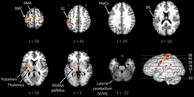
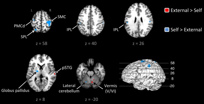
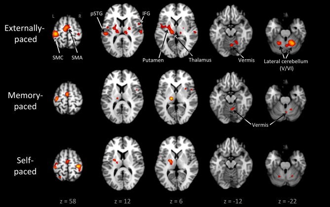
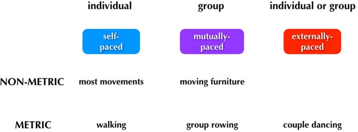
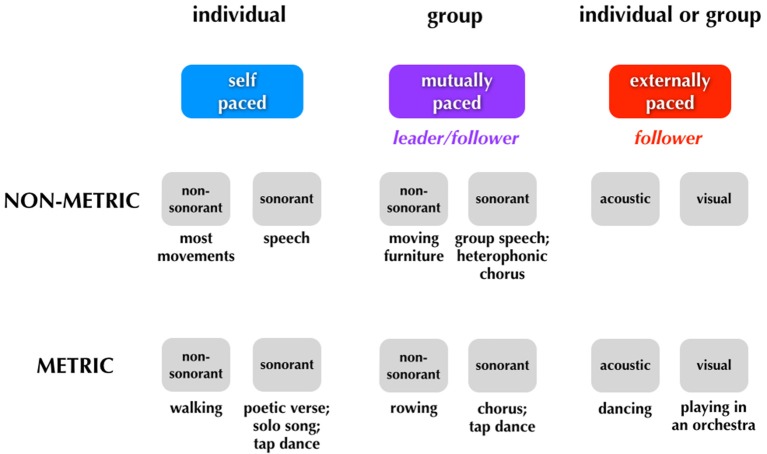
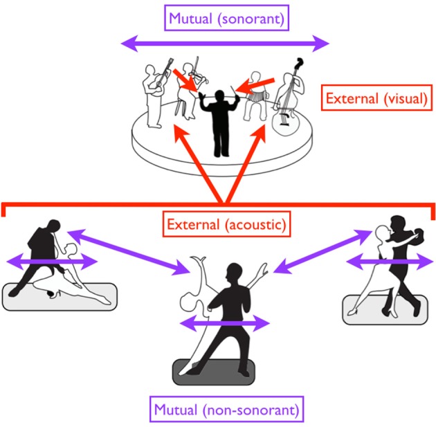
Synchronization of body movement to an acoustic rhythm is a major form of entrainment, such as occurs in dance. This is exemplified in experimental studies of finger tapping. Entrainment to a beat is contrasted with movement that is internally driven and is therefore self-paced. In order to examine brain areas important for entrainment to an acoustic beat, we meta-analyzed the functional neuroimaging literature on finger tapping (43 studies) using activation likelihood estimation (ALE) meta-analysis with a focus on the contrast between externally-paced and self-paced tapping. The results demonstrated a dissociation between two subcortical systems involved in timing, namely the cerebellum and the basal ganglia. Externally-paced tapping highlighted the importance of the spinocerebellum, most especially the vermis, which was not activated at all by self-paced tapping. In contrast, the basal ganglia, including the putamen and globus pallidus, were active during both types of tapping, but preferentially during self-paced tapping. These results suggest a central role for the spinocerebellum in audiomotor entrainment. We conclude with a theoretical discussion about the various forms of entrainment in humans and other animals.
Keywords: ALE; acoustic; basal ganglia; cerebellum; entrainment; finger tapping; meter; timing.
Figures
Similar articles
-
From Sound to Movement: Mapping the Neural Mechanisms of Auditory-Motor Entrainment and Synchronization.Brain Sci. 2024 Oct 25;14(11):1063. doi: 10.3390/brainsci14111063. Brain Sci. 2024. PMID: 39595826 Free PMC article. Review.
-
A functional MRI study of motor dysfunction in Friedreich's ataxia.Brain Res. 2012 Aug 30;1471:138-54. doi: 10.1016/j.brainres.2012.06.035. Epub 2012 Jul 3. Brain Res. 2012. PMID: 22771856
-
Steady-state evoked potentials distinguish brain mechanisms of self-paced versus synchronization finger tapping.Hum Mov Sci. 2018 Oct;61:151-166. doi: 10.1016/j.humov.2018.07.007. Epub 2018 Aug 8. Hum Mov Sci. 2018. PMID: 30098488
-
A functional magnetic resonance imaging study of paced finger tapping in children.Pediatr Neurol. 2003 Feb;28(2):89-95. doi: 10.1016/s0887-8994(02)00492-7. Pediatr Neurol. 2003. PMID: 12699857
-
Neural substrates of internally-based and externally-cued timing: An activation likelihood estimation (ALE) meta-analysis of fMRI studies.Neurosci Biobehav Rev. 2019 Jan;96:197-209. doi: 10.1016/j.neubiorev.2018.10.003. Epub 2018 Oct 11. Neurosci Biobehav Rev. 2019. PMID: 30316722
Cited by
-
Dancers' Somatic of Musicality.Front Psychol. 2019 Dec 5;10:2681. doi: 10.3389/fpsyg.2019.02681. eCollection 2019. Front Psychol. 2019. PMID: 31866897 Free PMC article.
-
Autonomous Control of Music to Retrain Walking After Stroke.Neurorehabil Neural Repair. 2023 May;37(5):255-265. doi: 10.1177/15459683231174223. Epub 2023 Jun 5. Neurorehabil Neural Repair. 2023. PMID: 37272500 Free PMC article.
-
Functional remapping in networks of the Parkinsonian brain: A preclinical neuroimaging perspective with clinical correlates.Transl Neurosci. 2025 Jun 14;16(1):20250374. doi: 10.1515/tnsci-2025-0374. eCollection 2025 Jan 1. Transl Neurosci. 2025. PMID: 40574753 Free PMC article. Review.
-
Auditory Cueing of Pre-Learned Skills and Role of Subcortical Information Processing to Maximize Rehabilitative Outcomes Bridging Science and Music-Based Interventions.Healthcare (Basel). 2022 Nov 3;10(11):2207. doi: 10.3390/healthcare10112207. Healthcare (Basel). 2022. PMID: 36360548 Free PMC article.
-
Musical Training Facilitates Exogenous Temporal Attention via Delta Phase Entrainment within a Sensorimotor Network.J Neurosci. 2023 May 3;43(18):3365-3378. doi: 10.1523/JNEUROSCI.0220-22.2023. Epub 2023 Mar 28. J Neurosci. 2023. PMID: 36977585 Free PMC article. Clinical Trial.
References
LinkOut - more resources
Full Text Sources
Other Literature Sources
Miscellaneous