Review
. 2014 Sep 30:5:1040.
doi: 10.3389/fpsyg.2014.01040. eCollection 2014.

A healthy heart is not a metronome: an integrative review of the heart's anatomy and heart rate variability

Affiliations
Review

A healthy heart is not a metronome: an integrative review of the heart's anatomy and heart rate variability

Fred Shaffer et al. Front Psychol. .

Abstract

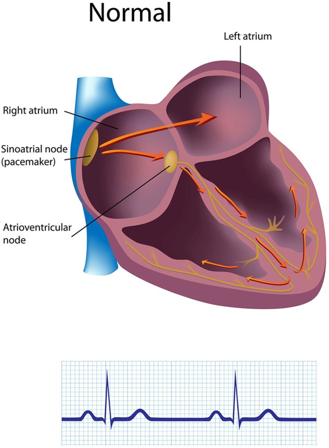
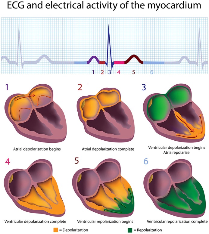
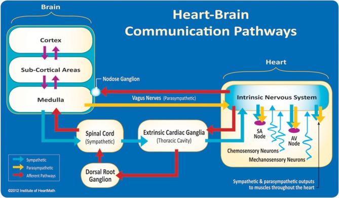
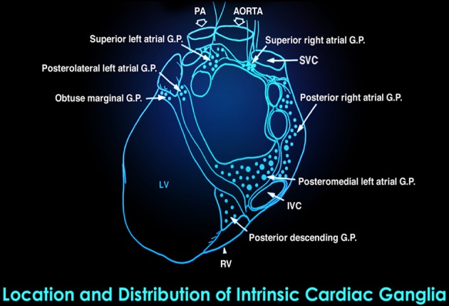
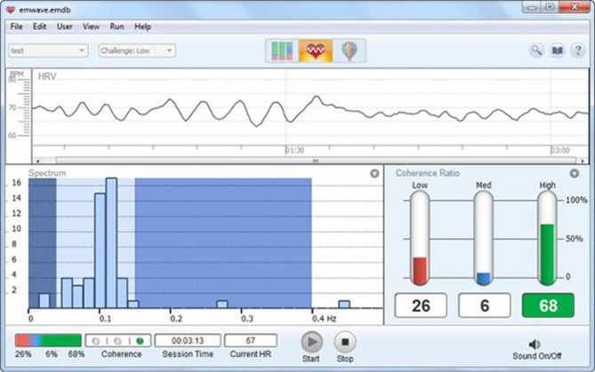
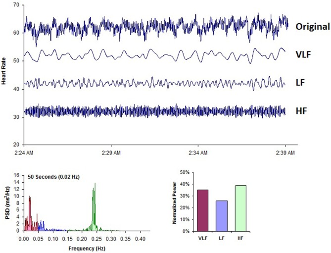
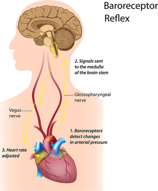
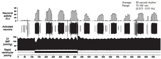
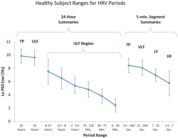
Heart rate variability (HRV), the change in the time intervals between adjacent heartbeats, is an emergent property of interdependent regulatory systems that operate on different time scales to adapt to challenges and achieve optimal performance. This article briefly reviews neural regulation of the heart, and its basic anatomy, the cardiac cycle, and the sinoatrial and atrioventricular pacemakers. The cardiovascular regulation center in the medulla integrates sensory information and input from higher brain centers, and afferent cardiovascular system inputs to adjust heart rate and blood pressure via sympathetic and parasympathetic efferent pathways. This article reviews sympathetic and parasympathetic influences on the heart, and examines the interpretation of HRV and the association between reduced HRV, risk of disease and mortality, and the loss of regulatory capacity. This article also discusses the intrinsic cardiac nervous system and the heart-brain connection, through which afferent information can influence activity in the subcortical and frontocortical areas, and motor cortex. It also considers new perspectives on the putative underlying physiological mechanisms and properties of the ultra-low-frequency (ULF), very-low-frequency (VLF), low-frequency (LF), and high-frequency (HF) bands. Additionally, it reviews the most common time and frequency domain measurements as well as standardized data collection protocols. In its final section, this article integrates Porges' polyvagal theory, Thayer and colleagues' neurovisceral integration model, Lehrer et al.'s resonance frequency model, and the Institute of HeartMath's coherence model. The authors conclude that a coherent heart is not a metronome because its rhythms are characterized by both complexity and stability over longer time scales. Future research should expand understanding of how the heart and its intrinsic nervous system influence the brain.

Keywords: biofeedback interventions; emotional self-regulation; heart rate variability; neurocardiology; psychophysiological coherence.


Figures

Figure 1
Figure 1
The generation of the electrocardiogram. Credit: Alila Sao Mai/Shutterstock.com.
Figure 2
Figure 2
The depolarization and repolarization of the heart. Credit: Alila Sao Mai/Shutterstock.com.
Figure 3
Figure 3
The neural communication pathways interacting between the heart and the brain are responsible for the generation of HRV. The intrinsic cardiac nervous system integrates information from the extrinsic nervous system and from the sensory neurites within the heart. The extrinsic cardiac ganglia located in the thoracic cavity have connections to the lungs and esophagus and are indirectly connected via the spinal cord to many other organs such as the skin and arteries. The vagus nerve (parasympathetic) primarily consists of afferent (flowing to the brain) fibers which connect to the medulla, after passing through the nodose ganglion. Credit: Institute of HeartMath.
Figure 4
Figure 4
Microscopic image of interconnected intrinsic cardiac ganglia in the human heart. The thin, light blue structures are multiple axons that connect the ganglia. Credit: Dr. Andrew Armour and the Institute of HeartMath.
Figure 5
Figure 5
This drawing shows the location and distribution of intrinsic cardiac ganglia which are interconnected and form the “heart brain.” Note how they are distributed around the orifices of the major vessels. Credit: Dr. Andrew Armour and the Institute of HeartMath.
Figure 6
Figure 6
Display of short-term HRV activity. Credit: Institute of HeartMath.
Figure 7
Figure 7
ECG electrode placement. Credit: Truman State University Center for Applied Psychophysiology.
Figure 8
Figure 8
This figure shows a typical HRV recording over a 15-min period during resting conditions in a healthy individual. The top trace shows the original HRV waveform. Filtering techniques were used to separate the original waveform into VLF, LF, and HF bands as shown in the lower traces. The bottom of the figure shows the power spectra (left) and the percentage of power (right) in each band. Credit: Institute of HeartMath.
Figure 9
Figure 9
Credit: Alila Sao Mai/Shutterstock.com.
Figure 10
Figure 10
Long-term single-neuron recordings from an afferent neuron in the intrinsic cardiac nervous system in a beating dog heart. The top row shows neural activity, the second row, the actual neural recording, and the third row, the left ventricular pressure. This intrinsic rhythm has an average period of 90 s with a range between 75 and 100 s (0.013–0.01 Hz), which falls within the VLF band. Credit: Dr. Andrew Armour and the Institute of HeartMath.
Figure 11
Figure 11
This figure shows the power in the various frequency bands for 24-h HRV and 95% confidence intervals for each of the bands. The left side of the figure reveals a number of slower rhythms that make up the ULF band. The analysis was conducted using the healthy sample described in Umetani et al. (1998). The right side of the figure shows an analysis of the same data performed on 5-min segments as is traditionally done. Credit: Institute of HeartMath.

References

    1. Agelink M. W., Boz C., Ullrich H., Andrich J. (2002). Relationship between major depression and heart rate variability. Clinical consequences and implications for antidepressive treatment. Psychiatry Res. 113, 139–149 10.1016/S0165-1781(02)00225-1 - DOI - PubMed
    1. Ahmed A. K., Harness J. B., Mearns A. J. (1982). Respiratory control of heart rate. Eur. J. Appl. Physiol. 50, 95–104 10.1007/BF00952248 - DOI
    1. Akselrod S., Gordon D., Ubel F. A., Shannon D. C., Barger A. C., Cohen R. J. (1981). Power spectrum analysis of heart rate fluctuation: a quantitative probe of beat-to-beat cardiovascular control. Science 213, 220–222 10.1126/science.6166045 - DOI - PubMed
    1. Alabdulgader A. A. (2012). Coherence: a novel nonpharmacological modality for lowering blood pressure in hypertensive patients. Glob. Adv. Health Med. 1, 56–64 10.7453/gahmj.2012.1.2.011 - DOI - PMC - PubMed
    1. Ardell J. L., Butler C. K., Smith F. M., Hopkins D. A., Armour J. A. (1991). Activity of in vivo atrial and ventricular neurons in chronically decentralized canine hearts. Am. J. Physiol. 260, H713–H721 - PubMed