

Hydrocephalus and neurocysticercosis: cases illustrative of three distinct mechanisms
- PMID: 25324888
- PMCID: PMC4198720
- DOI: 10.3988/jcn.2014.10.4.363
Hydrocephalus and neurocysticercosis: cases illustrative of three distinct mechanisms
Abstract
Background: Cysticercosis is the most frequent parasitic infection of the nervous system. Most lesions are intracranial, and spinal involvement is rare. We describe here in two cases of neurocysticercosis (NCC) in the brain and one in the spinal cord that illustrate three distinct mechanisms leading to symptomatic acute hydrocephalus.
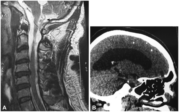
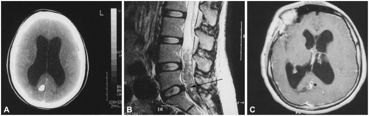
Case report: Hydrocephalus was related to intracranial NCC in two of them. In the first case the hydrocephalus was due to an extensive arachnoiditis to the craniocervical junction, while in the second it was caused by obstruction of Magendie's foramen in the fourth ventricle by the scolex of Taenia solium. For the third patient, hydrocephalus revealed cysticercosis of the cauda equina due to the scolex.
Conclusions: NCC should be considered as a possible diagnosis for patients suffering from hydrocephalus when they originate from or have traveled in endemic areas, MRI of the spine is mandatory to search for intraspinal lesions.
Keywords: brain; cauda equina; hydrocephalus; neurocysticercosis; tropical disease.
Conflict of interest statement
The authors have no financial conflicts of interest.
Figures

Similar articles
-
[Treatment of racemose neurocysticercosis of the spine].Rev Neurol. 2005 May 1-15;40(9):544-7. Rev Neurol. 2005. PMID: 15898016 Spanish.
-
Hydrocephalus due to Membranous Obstruction of Magendie's Foramen.J Korean Neurosurg Soc. 2015 Jan;57(1):68-71. doi: 10.3340/jkns.2015.57.1.68. Epub 2015 Jan 31. J Korean Neurosurg Soc. 2015. PMID: 25674349 Free PMC article.
-
"Malignant" Craniospinal Neurocysticercosis: A Rare Case.World Neurosurg. 2021 Feb;146:95-102. doi: 10.1016/j.wneu.2020.10.121. Epub 2020 Oct 27. World Neurosurg. 2021. PMID: 33127574
-
Hydrocephalus in neurocysticercosis.Childs Nerv Syst. 2011 Oct;27(10):1709-21. doi: 10.1007/s00381-011-1500-3. Epub 2011 Sep 17. Childs Nerv Syst. 2011. PMID: 21928035 Review.
-
Clinical manifestations, diagnosis, and treatment of neurocysticercosis.Curr Neurol Neurosci Rep. 2011 Dec;11(6):529-35. doi: 10.1007/s11910-011-0226-7. Curr Neurol Neurosci Rep. 2011. PMID: 21915772 Review.
Cited by
-
An Unusual Presentation of Neurocysticercosis: A Space-Occupying Lesion in the Fourth Ventricle Associated with Progressive Cognitive Decline.Am J Trop Med Hyg. 2016 Jan;94(1):172-5. doi: 10.4269/ajtmh.15-0099. Epub 2015 Nov 30. Am J Trop Med Hyg. 2016. PMID: 26621562 Free PMC article.
-
The leptomeninges as a critical organ for normal CNS development and function: First patient and public involved systematic review of arachnoiditis (chronic meningitis).PLoS One. 2022 Sep 30;17(9):e0274634. doi: 10.1371/journal.pone.0274634. eCollection 2022. PLoS One. 2022. PMID: 36178925 Free PMC article.
-
Isolated Intramedullary Lumbar Spine Neurocysticercosis: A Rare Occurrence and Review of Literature.Surg J (N Y). 2021 Dec 15;7(4):e327-e336. doi: 10.1055/s-0041-1739118. eCollection 2021 Oct. Surg J (N Y). 2021. PMID: 34926816 Free PMC article.
-
Brainstem and Spinal Arachnoiditis Ossificans Associated With Neurocysticercosis: A Case Report.Cureus. 2023 Sep 28;15(9):e46157. doi: 10.7759/cureus.46157. eCollection 2023 Sep. Cureus. 2023. PMID: 37905247 Free PMC article.
-
Human Neurocysticercosis: An Overview.Pathogens. 2022 Oct 20;11(10):1212. doi: 10.3390/pathogens11101212. Pathogens. 2022. PMID: 36297269 Free PMC article. Review.
References
-
- Lobato RD, Lamas E, Portillo JM, Roger R, Esparza J, Rivas JJ, et al. Hydrocephalus in cerebral cysticercosis. Pathogenic and therapeutic considerations. J Neurosurg. 1981;55:786–793. - PubMed
-
- Raccurt CP, Agnamey P, Boncy J, Henrys JH, Totet A. Seroprevalence of human Taenia solium cysticercosis in Haiti. J Helminthol. 2009;83:113–116. - PubMed
-
- Bouree P, Dumazedier D, Bisaro F, Resende P, Comoy J, Aghakhani N. Spinal cord cysticercosis: a case report. J Egypt Soc Parasitol. 2006;36:727–736. - PubMed
-
- Monteiro L, Almeida-Pinto J, Stocker A, Sampaio-Silva M. Active neurocysticercosis, parenchymal and extraparenchymal: a study of 38 patients. J Neurol. 1993;241:15–21. - PubMed
LinkOut - more resources
Full Text Sources
Other Literature Sources
