

doi: 10.3315/jdcr.2014.1177.
Photoletter to the editor: Collision tumor of melanoma and atypical fibroxanthoma of the scalp
Affiliations
- PMID: 25324912
- PMCID: PMC4195507
- DOI: 10.3315/jdcr.2014.1177
Item in Clipboard
Photoletter to the editor: Collision tumor of melanoma and atypical fibroxanthoma of the scalp
J Dermatol Case Rep.
.
Abstract
Several combinations of different skin tumors occuring one adjacent to the other or even in a single lesion have been described up to date. Collision tumors involving atypical fibroxanthoma and melanoma are extremely uncommon. Herein we present a case of melanoma associated with AFX and discuss on the usefulness of dermoscopy in the clinical diagnosis of collision tumors.
Keywords: collision tumor; dermoscopy; fibroxanthoma; melanoma; skin cancer; trichoscopy.
Figures
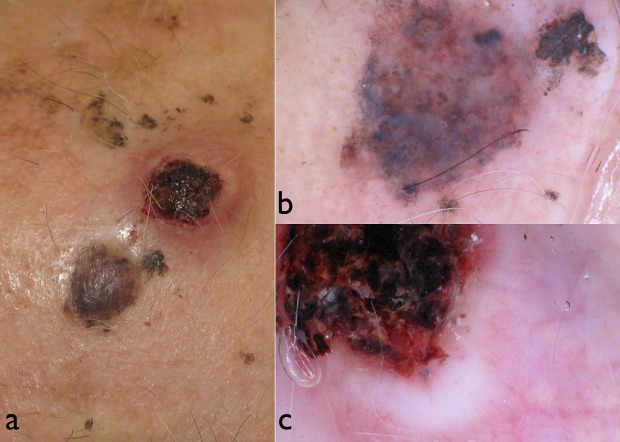
(a) Clinical aspect of a pigmented and an ulcerated nodule developing contiguously on the scalp. (b) Dermoscopy of the pigmented nodule revealed irregular blotches, irregular globules, a blue-white veil and remnants of pigment network at the periphery, overall suggestive of melanoma. (c) A large ulceration, whitish-pinkish areas and a polymorphous vascular pattern dermoscopically characterized the non-pigmented nodule.
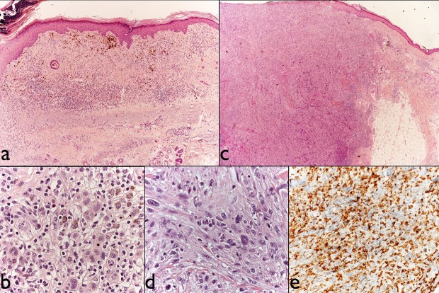
(a) Melanoma. Histopathologic examination reveals a dermal-based proliferation of atypical pigmented melanocytes on a background of severely damaged skin. The inflammatory infiltrate and the superficial erosion in the left upper corner are the residual sign of the previous punch biopsy. (b) Melanoma cells at higher magnification. (c) Atypical fibroxanthoma. On histology, the dermis is diffusely infiltrated by a richly cellular neoplasm. (d) At higher magnification, it is composed of pleomorphic and mitotically active spindle cells. (e) The neoplastic cells are diffusely immunoreactive with CD68.
Similar articles
-
Dermoscopy features of atypical fibroxanthoma: A multicenter study of the International Dermoscopy Society.Australas J Dermatol. 2018 Nov;59(4):309-314. doi: 10.1111/ajd.12802. Epub 2018 Mar 23. Australas J Dermatol. 2018. PMID: 29569417
-
Collision Tumor Comprised of Atypical Fibroxanthoma and Invasive Squamous Cell Carcinoma: A Case Report of an Extremely Rare Entity.Cureus. 2022 Sep 19;14(9):e29324. doi: 10.7759/cureus.29324. eCollection 2022 Sep. Cureus. 2022. PMID: 36277592 Free PMC article.
-
Clear cell atypical fibroxanthoma: a case report and review of the literature.J Cutan Pathol. 2016 Jun;43(6):538-542. doi: 10.1111/cup.12696. Epub 2016 Apr 5. J Cutan Pathol. 2016. PMID: 26956561
-
Atypical Fibroxanthoma - Histological Diagnosis, Immunohistochemical Markers and Concepts of Therapy.Anticancer Res. 2015 Nov;35(11):5717-35. Anticancer Res. 2015. PMID: 26503993 Review.
-
[Atypical fibroxanthoma of the scalp: overview and recent developments].Hautarzt. 2014 Dec;65(12):1008-10. doi: 10.1007/s00105-014-3541-5. Hautarzt. 2014. PMID: 25392128 Review. German.
Cited by
-
CD10 and p63 expression in a sarcomatoid undifferentiated melanoma: A cautionary (and molecularly annotated) tale.J Cutan Pathol. 2020 Jun;47(6):541-547. doi: 10.1111/cup.13646. Epub 2020 Jan 20. J Cutan Pathol. 2020. PMID: 31943331 Free PMC article.
-
Collision skin lesions-results of a multicenter study of the International Dermoscopy Society (IDS).Dermatol Pract Concept. 2017 Jul 31;7(4):51-62. doi: 10.5826/dpc.0704a12. eCollection 2017 Oct. Dermatol Pract Concept. 2017. PMID: 29230351 Free PMC article.
-
Atypical fibroxanthoma†.J Surg Case Rep. 2015 Mar 4;2015(3):rjv010. doi: 10.1093/jscr/rjv010. J Surg Case Rep. 2015. PMID: 25742967 Free PMC article.
References
-
- Lallas A, Moscarella E, Argenziano G, Longo C, Apalla Z, Ferrara G, Piana S, Rosato S, Zalaudek I. Dermoscopy of uncommon skin tumours. Australas J Dermatol. 2014;55:53–62. - PubMed
-
- Zaballos P, Llambrich A, Puig S, Malvehy J. Dermoscopy is useful for the recognition of benign-malignant compound tumours. Br J Dermatol. 2005;153:653–656. - PubMed
-
- McGregor DH, Cherian R, Romanas MM, Ulusarac O, Mathur SC, Feldman MM. Amelanotic malignant melanoma: two collision tumors presenting as basal cell carcinoma and atypical fibroxanthoma. Ann Clin Lab Sci. 2008 Spring;38:157–162. - PubMed
-
- Wilsher MJ. Collision tumour: atypical fibroxanthoma and invasive melanoma. Pathology. 2009;41:699–701. - PubMed
-
- Alves R, Ocaña J, Vale E, Correia S, Viana I, Bordalo O. Basal cell carcinoma and atypical fibroxanthoma: an unusual collision tumor. J Am Acad Dermatol. 2010;63:e74–76. - PubMed
LinkOut - more resources
Full Text Sources
Other Literature Sources