. 2014 Sep 29:4:251.
doi: 10.3389/fonc.2014.00251. eCollection 2014.

Hidden treasures in "ancient" microarrays: gene-expression portrays biology and potential resistance pathways of major lung cancer subtypes and normal tissue

Affiliations

Hidden treasures in "ancient" microarrays: gene-expression portrays biology and potential resistance pathways of major lung cancer subtypes and normal tissue

Konstantinos Kerkentzes et al. Front Oncol. .

Abstract

Objective: Novel statistical methods and increasingly more accurate gene annotations can transform "old" biological data into a renewed source of knowledge with potential clinical relevance. Here, we provide an in silico proof-of-concept by extracting novel information from a high-quality mRNA expression dataset, originally published in 2001, using state-of-the-art bioinformatics approaches.

Methods: The dataset consists of histologically defined cases of lung adenocarcinoma (AD), squamous (SQ) cell carcinoma, small-cell lung cancer, carcinoid, metastasis (breast and colon AD), and normal lung specimens (203 samples in total). A battery of statistical tests was used for identifying differential gene expressions, diagnostic and prognostic genes, enriched gene ontologies, and signaling pathways.

Results: Our results showed that gene expressions faithfully recapitulate immunohistochemical subtype markers, as chromogranin A in carcinoids, cytokeratin 5, p63 in SQ, and TTF1 in non-squamous types. Moreover, biological information with putative clinical relevance was revealed as potentially novel diagnostic genes for each subtype with specificity 93-100% (AUC = 0.93-1.00). Cancer subtypes were characterized by (a) differential expression of treatment target genes as TYMS, HER2, and HER3 and (b) overrepresentation of treatment-related pathways like cell cycle, DNA repair, and ERBB pathways. The vascular smooth muscle contraction, leukocyte trans-endothelial migration, and actin cytoskeleton pathways were overexpressed in normal tissue.

Conclusion: Reanalysis of this public dataset displayed the known biological features of lung cancer subtypes and revealed novel pathways of potentially clinical importance. The findings also support our hypothesis that even old omics data of high quality can be a source of significant biological information when appropriate bioinformatics methods are used.

Keywords: bioinformatics; carcinoid; lung adenocarcinoma; mesothelioma; microarray; small cell; squamous.


Figures
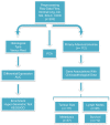
Figure 1
Figure 1
Flow-chart of the performed analyses. This figure illustrates the analyses performed during this study. The raw CEL files were obtained from the site of the authors of the original publication and were preprocessed with the RMA algorithm. Three different branches of analyses were performed; first, PCA was used for investigating the relationship among different types of tumors and against normal tissues. Second, differentially expressed and diagnostic genes were identified in each histological type versus the rest. On the basis of these lists of differentially expressed genes, enriched GO biological processes, and KEGG pathways were identified with the hyper-geometric test. Finally, the association between clinicopathological and transcriptional information were investigated in the primary adenocarcinoma samples. We tried to identify genes correlated with the tumor size, differentially expressed and diagnostic genes of lymph node status and metastasis, and single gene or gene signatures predictive of survival. The sample size differs because complete clinicopathological information was not always provided and in the survival analysis only the primary adenocarcinomas were taken into account.
Figure 2
Figure 2
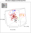
Distribution of normal tissues and cancer subtypes in the principal component space. This visualization shows that normal and carcinoids (COID) have distinct expression profiles, while adenocarcinomas (AD) overlap with both squamous (SQ) and small-cell carcinoma (SCLC).
Figure 3
Figure 3
Distribution of normal tissues and cancer subtypes except adenocarcinoma in the principal component space. Small-cell, squamous, and carcinoid subgroups show distinctive expression profile when the adenocarcinomas are omitted, indicating that adenocarcinoma is the most heterogeneous group.
Figure 4
Figure 4
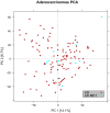
Distribution of primary versus metastatic adenocarcinoma samples in the principal component space. PCA plot showing the expression of the adenocarcinoma metastases to the lung from other locations (light blue) in relation to primary lung cancers including adenocarcinoma (red). The general expression profile of the adenocarcinoma metastases versus primary adenocarcinomas did not vary significantly.
Figure 5
Figure 5
Comparison of protein and gene-expression patterns in primary lung neoplasias. (A) hematoxylin and eosin (H&E), (B) thyroid transcription factor-1 (TTF1), (C) cytokeratin 5 (CK5), (D) p63, and (E) synaptophysin (SNP). All photos ×200. (1) Adenocarcinoma, (A) H&E showing low differentiation with solid growth, (B) TTF1 showing strong nuclear staining of all tumor cells, (C) CK5 showing no cytoplasmic staining of tumor cells, (D) p63 showing nuclear staining of a few scattered tumor cells, and (E) SNP showing cytoplasmic staining of a few scattered tumor cells. (2) Squamous cell carcinoma, (A) H&E showing solid growth with slight squamous maturation to the left, (B) TTF1 showing no nuclear staining of tumor cells, scattered entrapped alveolar cells with normal TTF1 expression is seen, (C) CK5 showing strong cytoplasmic staining of most tumor cells, (E) p63 showing widespread nuclear staining of tumor cells, in the upper right corner an entrapped bronchiolus with normal p63 expression of basal cells is seen, and (E) SNP showing no cytoplasmic staining of tumor cells. (3) Small-cell carcinoma, (A) H&E showing sheet-like growth, (B) TTF1 showing very strong nuclear staining of tumor cells, (C) CK5 showing no staining of tumor cells, (D) p63 showing widespread but weak nuclear staining of tumor cells, and (E) SNP showing strong cytoplasmic staining of tumor cells. (4) Carcinoid, (A) H&E showing trabecular growth, (B) TTF1 showing moderate nuclear staining of most tumor cells, (C) CK5 showing no cytoplasmic staining of tumor cells, (D) p63 showing no nuclear staining of tumor cells; a few entrapped alveolar cells are positive, and (E) SNP showing strong cytoplasmic staining of tumor cells.
Figure 6
Figure 6
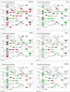
Cell cycle pathway. Tumor suppressor genes (CDKN1A/p21, CDKN1C/p57, RBL2/p130) and the cyclin D family were overexpressed in normal lung tissue while there was type-specific overexpression of oncogenes, cyclins, cyclin dependent kinases, and other cell cycle driving genes in tumors. Genes belonging to the mini-chromosome maintenance complex were overexpressed in squamous and small-cell lung cancer, as well as in mesothelioma. The same genes were not differentially expressed in AD and downregulated in COID. Fifty-six out of 63 genes in the Cell Cycle pathway were overexpressed in SQ, including CCNE/CDK2, CCNA/CDK2, CCNA/CDK1, CCNB/CDK1 complexes, BUB1, BUB1B, BUB3, and MYC oncogenes, damage response genes (ATR, CHEK1/2) and five genes of the 14-3-3 family. Red is overexpressed, dark green downregulated. SQ, squamous type; AD, adenocarcinoma; SCLC, small-cell lung cancer; COID, carcinoid; MESO, mesothelioma.
Figure 7
Figure 7
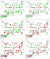
Nucleotide excision repair (NER) pathway. Genes belonging to all the principal DNA repair systems were predominantly overexpressed in tumors. The NER is the most analyzed pathway related to cisplatin resistance. In normal lung tissue (Normal), only one DNA repair gene was overexpressed, the XPC. In the COID, SCLC, SQ, and MESO tumors, an array of genes was overexpressed. Key genes of the whole repair process were overexpressed. In the SQ, 25 NER genes were differentially expressed, and only four were downregulated, the CUL4, XPD, ERCC2, and ERCC8. The most heterogeneous tumor group, AD, had the least NER genes overexpressed. Red indicates significantly overexpression, dark green significant downregulation, and light green no differential expression. COID, carcinoids; AD, adenocarcinoma; SCLC, small-cell lung cancer; SQ, Squamous cell lung cancer; MESO, mesothelioma.
Figure 8
Figure 8
Homologous recombination (HR) pathway. The HR is the principal, non-error-prone, repair mechanism for DNA double-strand breaks. SQ and SCLC had 10 of 11 overexpressed genes throughout the pathway, including the very important BRCA2, RAD51 paralogs, and BLM (Bloom syndrome mutated). Red indicates significantly overexpression, dark green significant downregulation, and light green no differential expression. COID, carcinoids; AD, adenocarcinoma; SCLC, small-cell lung cancer; SQ, Squamous cell lung cancer; MESO, mesothelioma.
Figure 9
Figure 9
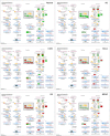
Vascular smooth muscle contraction pathway. Genes belonging to the vascular smooth muscle contraction pathway were significantly overrepresented and upregulated in normal tissue (15/36 genes, p < 0.0001). None of the genes of the smooth muscle cell membrane (according to KEGG pathways) were overexpressed in SQ, SCLC, and mesothelioma. Red indicates significantly overexpression, dark green significant downregulation, and light green no differential expression. COID, carcinoids; AD, adenocarcinoma; SCLC, small- cell lung cancer; SQ, squamous cell lung cancer; MESO, mesothelioma.

References

    1. Alberg AJ, Brock MV, Ford JG, Samet JM, Spivack SD. Epidemiology of lung cancer: diagnosis and management of lung cancer, 3rd ed: American College of Chest Physicians evidence-based clinical practice guidelines. Chest (2013) 143:1S–29S10.1378/chest.12-2345 - DOI - PMC - PubMed
    1. Mirsadraee S, Oswal D, Alizadeh Y, Caulo A, van Beek E., Jr The 7th lung cancer TNM classification and staging system: review of the changes and implications. World J Radiol (2012) 4:128–3410.4329/wjr.v4.i4.128 - DOI - PMC - PubMed
    1. Travis WD, Brambilla E, Riely GJ. New pathologic classification of lung cancer: relevance for clinical practice and clinical trials. J Clin Oncol (2013) 31:992–100110.1200/JCO.2012.46.9270 - DOI - PubMed
    1. Smit EF, Socinski MA, Mullaney BP, Myrand SP, Scagliotti GV, Lorigan P, et al. Biomarker analysis in a phase III study of pemetrexed-carboplatin versus etoposide-carboplatin in chemonaive patients with extensive-stage small-cell lung cancer. Ann Oncol (2012) 23:1723–910.1093/annonc/mdr563 - DOI - PubMed
    1. Subramanian J, Govindan R. Chemotherapy: continued lack of progress in SCLC. Nat Rev Clin Oncol (2010) 7:77–810.1038/nrclinonc.2009.223 - DOI - PubMed