

Postsynaptic inhibition of hypoglossal motoneurons produces atonia of the genioglossal muscle during rapid eye movement sleep
- PMID: 25325470
- PMCID: PMC4262947
- DOI: 10.5665/sleep.4340
Postsynaptic inhibition of hypoglossal motoneurons produces atonia of the genioglossal muscle during rapid eye movement sleep
Abstract
Study objectives: Hypoglossal motoneurons were recorded intracellularly to determine whether postsynaptic inhibition or disfacilitation was responsible for atonia of the lingual muscles during rapid eye movement (REM) sleep.
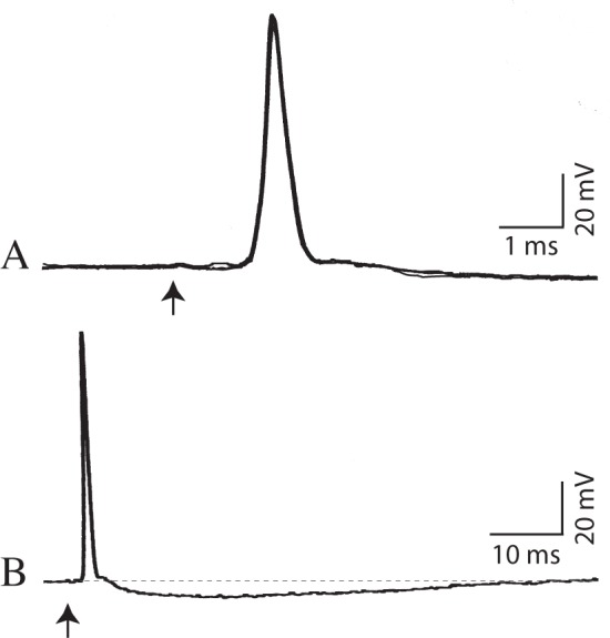
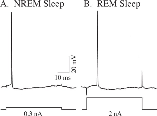
Design: Intracellular records were obtained of the action potentials and subthreshold membrane potential activity of antidromically identified hypoglossal motoneurons in cats during wakefulness, nonrapid eye movement (NREM) sleep, and REM sleep. A cuff electrode was placed around the hypoglossal nerve to antidromically activate hypoglossal motoneurons. The state-dependent changes in membrane potential, spontaneous discharge, postsynaptic potentials, and rheobase of hypoglossal motoneurons were determined.
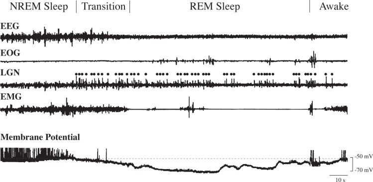
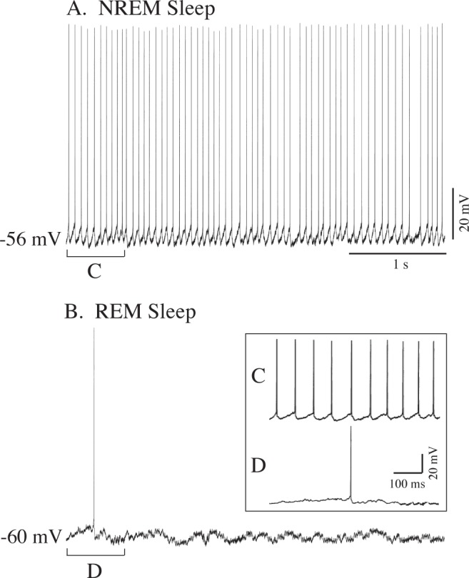
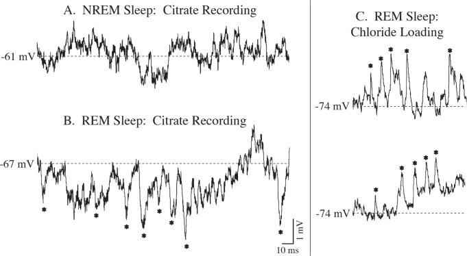
Analyses and results: During quiet wakefulness and NREM sleep, hypoglossal motoneurons exhibited spontaneous repetitive discharge. In the transition from NREM sleep to REM sleep, repetitive discharge ceased and the membrane potential began to hyperpolarize; maximal hyperpolarization (10.5 mV) persisted throughout REM sleep. During REM sleep there was a significant increase in rheobase, which was accompanied by barrages of large-amplitude inhibitory postsynaptic potentials (IPSPs), which were reversed following the intracellular injection of chloride ions. The latter result indicates that they were mediated by glycine; IPSPs were not present during wakefulness or NREM sleep.
Conclusions: We conclude that hypoglossal motoneurons are postsynaptically inhibited during naturally occurring REM sleep; no evidence of disfacilitation was observed. The data also indicate that glycine receptor-mediated postsynaptic inhibition of hypoglossal motoneurons is crucial in promoting atonia of the lingual muscles during REM sleep.
Keywords: IPSP; OSA; REM sleep; atonia; hypoglossal; motoneuron; postsynaptic inhibition.
© 2014 Associated Professional Sleep Societies, LLC.
Figures
Similar articles
-
Control of hypoglossal motoneurones during naturally occurring sleep and wakefulness in the intact, unanaesthetized cat: a field potential study.J Sleep Res. 2014 Aug;23(4):469-74. doi: 10.1111/jsr.12137. Epub 2014 Mar 8. J Sleep Res. 2014. PMID: 24605864
-
Suppression of hypoglossal motoneurons during the carbachol-induced atonia of REM sleep is not caused by fast synaptic inhibition.Brain Res. 1993 May 21;611(2):300-12. doi: 10.1016/0006-8993(93)90517-q. Brain Res. 1993. PMID: 8334524
-
Glycinergic and GABA(A)-mediated inhibition of somatic motoneurons does not mediate rapid eye movement sleep motor atonia.J Neurosci. 2008 Apr 2;28(14):3535-45. doi: 10.1523/JNEUROSCI.5023-07.2008. J Neurosci. 2008. PMID: 18385312 Free PMC article.
-
Synaptic mechanisms and circuitry involved in motoneuron control during sleep.Int Rev Neurobiol. 1983;24:213-58. doi: 10.1016/s0074-7742(08)60223-8. Int Rev Neurobiol. 1983. PMID: 6197386 Review.
-
Confirmation of the consensus that glycinergic postsynaptic inhibition is responsible for the atonia of REM sleep.Sleep. 2008 Nov;31(11):1487-91. doi: 10.1093/sleep/31.11.1487. Sleep. 2008. PMID: 19014068 Free PMC article.
Cited by
-
Computational model of brain-stem circuit for state-dependent control of hypoglossal motoneurons.J Neurophysiol. 2018 Jul 1;120(1):296-305. doi: 10.1152/jn.00728.2017. Epub 2018 Apr 4. J Neurophysiol. 2018. PMID: 29617218 Free PMC article.
-
Catecholaminergic A1/C1 neurons contribute to the maintenance of upper airway muscle tone but may not participate in NREM sleep-related depression of these muscles.Respir Physiol Neurobiol. 2017 Oct;244:41-50. doi: 10.1016/j.resp.2017.07.001. Epub 2017 Jul 12. Respir Physiol Neurobiol. 2017. PMID: 28711601 Free PMC article.
-
Optical and pharmacological manipulation of hypoglossal motor nucleus identifies differential effects of taltirelin on sleeping tonic motor activity and responsiveness.Sci Rep. 2023 Jul 29;13(1):12299. doi: 10.1038/s41598-023-39562-z. Sci Rep. 2023. PMID: 37516800 Free PMC article.
-
The anatomical, cellular and synaptic basis of motor atonia during rapid eye movement sleep.J Physiol. 2016 Oct 1;594(19):5391-414. doi: 10.1113/JP271324. Epub 2016 Jul 3. J Physiol. 2016. PMID: 27060683 Free PMC article. Review.
-
Homeostatic regulation through GABA and acetylcholine muscarinic receptors of motor trigeminal neurons following sleep deprivation.Brain Struct Funct. 2017 Sep;222(7):3163-3178. doi: 10.1007/s00429-017-1392-4. Epub 2017 Mar 15. Brain Struct Funct. 2017. PMID: 28299422 Free PMC article.
References
-
- Chase MH. Motor control during sleep and wakefulness: clarifying controversies and resolving paradoxes. Sleep Med Rev. 2013;17:299–312. - PubMed
-
- Funk GD, Zwicker JD, Selvaratnam R, Robinson DM. Noradrenergic modulation of hypoglossal motoneuron excitability: developmental and putative state-dependent mechanisms. Arch Ital Biol. 2011;149:426–53. - PubMed
-
- Horner RL. Neural control of the upper airway: integrative physiological mechanisms and relevance for sleep disordered breathing. Compr Physiol. 2012;2:479–535. - PubMed
-
- White DP, Younes MK. Obstructive sleep apnea. Compr Physiol. 2012;2:2541–94. - PubMed
-
- Sauerland EK, Harper RM. The human tongue during sleep: electromyographic activity of the genioglossus muscle. Exp Neurol. 1976;51:160–70. - PubMed
Publication types
MeSH terms
Substances
Grants and funding
LinkOut - more resources
Full Text Sources
Other Literature Sources
Miscellaneous
